Identify the highlighted section using the drop down below.
What structure of the electrical conduction system passes through the highlighted area?
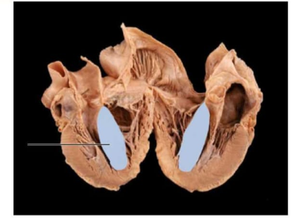
The Correct Answer is {"dropdown-group-1":"C","dropdown-group-2":"B"}
Correct answer:
- Interventricular septum
- Atrioventricular (AV) bundle / Bundle of His
The highlighted area represents the interventricular septum, the thick muscular wall that separates the right and left ventricles. It forms the medial wall of both ventricles and extends from the atrioventricular valves superiorly to the apex inferiorly. Its primary physiologic function is to prevent mixing of oxygenated blood in the left ventricle with deoxygenated blood in the right ventricle while also contributing to ventricular contraction. The electrical conduction structure that passes through this area is the atrioventricular (AV) bundle / Bundle of His. The Bundle of His passes from the atrioventricular node into the membranous portion of the interventricular septum. It then divides into the right and left bundle branches, which travel along the septum toward the apex to distribute electrical impulses to both ventricles. This allows coordinated ventricular depolarization and synchronized contraction.
Nursing Test Bank
Naxlex Comprehensive Predictor Exams
Related Questions
Correct Answer is A
Explanation
A. Z-line to Z-line: A sarcomere, the functional contractile unit of a cardiomyocyte, is defined as the region between two adjacent Z-lines (or Z-discs). The Z-lines anchor the thin filaments (actin) and mark the lateral boundaries of the sarcomere. During contraction, the sarcomere shortens as actin and myosin filaments slide past each other, generating tension and ultimately producing myocardial contraction.
B. A-line to A-line: The A-band corresponds to the length of the thick filaments (myosin) within a sarcomere and does not represent the full functional unit. It includes both overlapping regions with thin filaments and the central H-zone, but A-band boundaries do not define sarcomere length.
C. I-band to I-band: The I-band contains only thin filaments and is bisected by the Z-line. It shortens during contraction, but its boundaries alone do not encompass the entire sarcomere, making it an incomplete reference for the sarcomere’s limits.
D. M-line to M-line: The M-line lies at the center of the sarcomere, anchoring thick filaments. While it is important for structural integrity, using M-line to M-line does not define the full sarcomere; it represents only the midpoint rather than the full functional contractile unit.
Correct Answer is E
Explanation
A. Pulmonary artery: The pulmonary artery originates from the right ventricle and is positioned anterior to the ascending aorta as it exits the heart. Its physiologic role is to transport deoxygenated blood to the lungs for oxygenation. It is part of the pulmonary circulation and does not arise from or receive blood flow from the right coronary artery, which supplies myocardium.
B. Left anterior descending artery: The left anterior descending artery, also known as the anterior interventricular artery, branches from the left coronary artery and runs within the anterior interventricular sulcus toward the apex. It supplies the anterior wall of the left ventricle and the anterior two-thirds of the interventricular septum.
C. Circumflex artery: The circumflex artery arises from the left coronary artery and courses in the left atrioventricular (coronary) sulcus. It supplies the lateral and posterior portions of the left ventricle and may contribute to left atrial perfusion. Its anatomical origin from the left coronary artery excludes it from being a branch of the right coronary artery.
D. Anterior interventricular artery: The anterior interventricular artery lies in the anterior interventricular groove between the right and left ventricles. It provides blood supply to the interventricular septum and the anterior surfaces of both ventricles. As a branch of the left coronary artery, it does not represent a continuation of blood flow from the right coronary artery.
E. Right marginal artery: The right marginal artery is a direct branch of the right coronary artery and travels along the acute margin of the heart toward the apex. It supplies the right ventricular free wall and contributes to perfusion of the right myocardium. Its anatomical course and origin confirm that blood flows from the right coronary artery into the right marginal artery.
Whether you are a student looking to ace your exams or a practicing nurse seeking to enhance your expertise , our nursing education contents will empower you with the confidence and competence to make a difference in the lives of patients and become a respected leader in the healthcare field.
Visit Naxlex, invest in your future and unlock endless possibilities with our unparalleled nursing education contents today