Where are bronchovesicular breath sounds auscultated?
Over the major bronchi, where fewer alveoli are located
At the bases of the lungs, on the anterior and lateral thorax
Over the manubrium on the anterior thorax
Just above the clavicles on each side of the sternum
The Correct Answer is A
Choice A reason: Bronchovesicular breath sounds are intermediate sounds heard over the mainstem bronchi in the first and second intercostal spaces anteriorly and between the scapulae posteriorly. They reflect a mix of air moving through the larger airways and the surrounding lung tissue, characterized by equal inspiratory and expiratory durations and moderate pitch.
Choice B reason: The lung bases and lateral thoracic regions are the primary locations for auscultating vesicular breath sounds. These sounds are soft, low-pitched, and breezy, produced by air moving through smaller bronchioles and alveoli. In these areas, the inspiratory phase is significantly longer than the expiratory phase, which is often nearly silent.
Choice C reason: Sounds heard directly over the manubrium and the trachea are classified as bronchial or tracheal breath sounds. These are high-pitched, loud, and harsh in quality. In these locations, the expiratory phase is longer and more intense than the inspiratory phase, with a distinct pause or gap between the two cycles.
Choice D reason: The area just above the clavicles represents the lung apices. While breath sounds can be heard here, they are typically vesicular in nature unless there is underlying consolidation. Bronchovesicular sounds are specifically localized to the central chest areas where the bifurcation of the trachea into the primary bronchi occurs.
Nursing Test Bank
Naxlex Comprehensive Predictor Exams
Related Questions
Correct Answer is D
Explanation
Choice A reason: Palpating at the 5th intercostal space at the midclavicular line is the technique used to locate the apical pulse or point of maximal impulse. While this is an essential component of a comprehensive cardiovascular examination, it follows the assessment of the neck vessels rather than immediately succeeding the inspection of the carotid.
Choice B reason: A thrill is a palpable vibration that signifies turbulent blood flow; however, it is detected through palpation, not inspection. Inspection is limited to the visual observation of pulsations. One cannot "inspect" for a thrill, as it is a tactile finding that requires the nurse to place the pads of the fingers over the artery.
Choice C reason: Auscultation of the carotid artery for bruits is an important step, especially in older adults or those with suspected vascular disease. However, standard physical assessment sequences typically move from inspection to palpation before proceeding to auscultation. Palpation provides immediate data on the strength and rhythm of the pulse before listening for turbulence.
Choice D reason: Following the visual inspection of the carotid area for pulsations or masses, the nurse must palpate the arterial pulse. It is critical to palpate only one carotid artery at a time to avoid stimulating the baroreceptors in the carotid sinus, which could induce reflex bradycardia, syncope, or a dangerous reduction in cerebral blood flow.
Correct Answer is C
Explanation
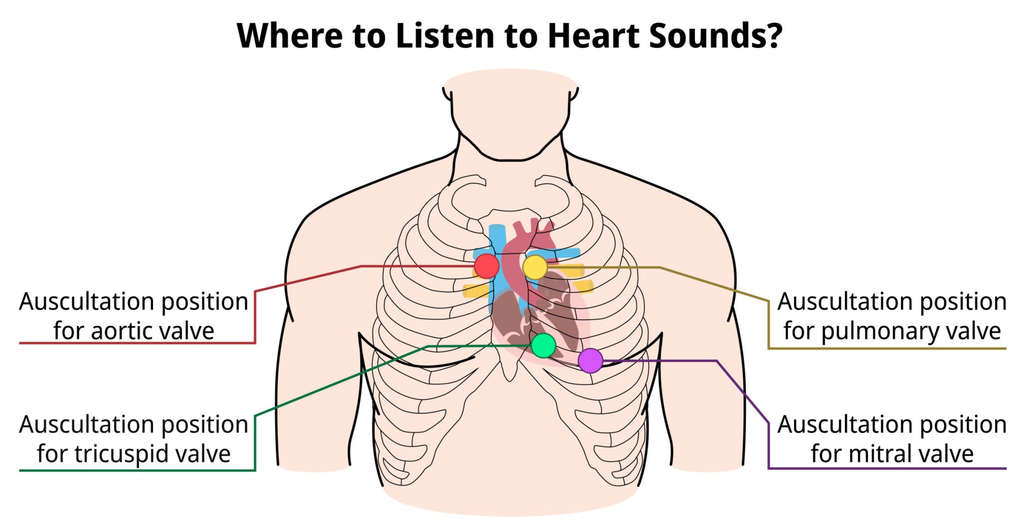
Choice A reason: The tricuspid valve is typically auscultated at the fourth or fifth intercostal space along the left lower sternal border. This anatomical site allows for the best transmission of sounds produced by the closure of the right atrioventricular valve, which separates the right atrium from the right ventricle during the cardiac cycle.
Choice B reason: The aortic valve is best assessed at the second intercostal space, just to the right of the sternal border. This location, known as the aortic area, facilitates the detection of the S2 heart sound and specific murmurs such as aortic stenosis, where blood flow is ejected into the ascending aorta.
Choice C reason: The mitral valve, or bicuspid valve, is located at the fifth intercostal space at the left midclavicular line, which corresponds to the cardiac apex. This site provides the most direct acoustic window to the left ventricle, making it the primary location for assessing the S1 heart sound and apical pulse.
Getty Images
Choice D reason: The pulmonic valve is auscultated at the second intercostal space, immediately to the left of the sternal border. This area is designated for monitoring the closure of the semilunar valve between the right ventricle and the pulmonary artery, where the pulmonic component of the second heart sound is loudest.
Whether you are a student looking to ace your exams or a practicing nurse seeking to enhance your expertise , our nursing education contents will empower you with the confidence and competence to make a difference in the lives of patients and become a respected leader in the healthcare field.
Visit Naxlex, invest in your future and unlock endless possibilities with our unparalleled nursing education contents today