Which letter represents ventricular depolarization?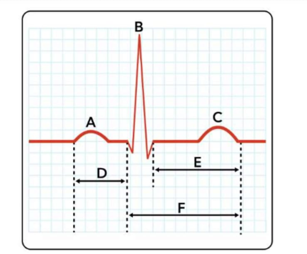
E
D
B
The Correct Answer is C
Correct answer: B.
B: Ventricular depolarization is the electrical activation of the ventricles that triggers ventricular contraction. It occurs after the impulse travels from the sinoatrial . On an electrocardiogram (ECG), ventricular depolarization is represented by the QRS complex, labelled as B in the diagram.
D: artrial depolarization - Atrial depolarization initiates atrial contraction, driven by impulses from the SA node. This electrical activity spreads across atrial myocardium, generating the P wave on the ECG and enabling ventricular filling.
E: ventricular repolarization - Ventricular repolarization restores the ventricles to their resting state after contraction. Represented by the T wave, it prepares the heart for the next depolarization, ensuring rhythmic cardiac cycles and effective pumping.
Nursing Test Bank
Naxlex Comprehensive Predictor Exams
Related Questions
Correct Answer is D
Explanation
A. It directly causes the initial calcium release from the sarcoplasmic reticulum: Calcium release from the sarcoplasmic reticulum is triggered by depolarization of the T-tubules and activation of ryanodine receptors, not directly by ATP. ATP provides energy for the mechanical steps of contraction but does not initiate calcium release.
B. It expands the H band by pushing thick filaments apart: The H band changes length passively as thin filaments slide over thick filaments during contraction. ATP does not mechanically push filaments apart; instead, it energizes myosin heads for cross-bridge cycling.
C. It prevents the troponin-tropomyosin complex from exposing binding sites: The troponin-tropomyosin complex blocks actin binding sites in the absence of calcium. ATP does not regulate this exposure; calcium binding to troponin shifts tropomyosin to allow myosin attachment.
D. It binds to myosin heads, allowing them to detach from actin: ATP binds to the myosin head after the power stroke, causing detachment from actin and breaking the actomyosin cross-bridge. ATP hydrolysis then re-cocks the myosin head, storing energy for the next contraction cycle. This is essential for continuous muscle contraction and relaxation in both skeletal and cardiac muscle.
Correct Answer is F
Explanation
Correct answer: F.
A. Trabeculae carneae: These are irregular, muscular ridges lining the inner walls of the ventricles. They prevent suction during contraction, aid in ventricular contraction efficiency, and contribute to overall cardiac structural integrity.
B. Pulmonary semilunar valve: This valve is located between the right ventricle and pulmonary artery. It prevents backflow of blood into the ventricle during diastole and ensures unidirectional pulmonary circulation toward the lungs.
C. Papillary muscles: Papillary muscles are conical projections of ventricular myocardium that anchor chordae tendineae. During ventricular contraction, they contract to prevent inversion or prolapse of atrioventricular valves, ensuring proper unidirectional blood flow.
D. Pectinate muscles: Pectinate muscles are comb-like muscular ridges in the atrial walls, particularly prominent in the right atrium. They enhance atrial contraction, increasing blood flow into the ventricles efficiently during systole.
E. Chordae tendineae: These are thin, fibrous cords connecting atrioventricular valve leaflets to papillary muscles. They prevent valve prolapse during ventricular contraction, maintaining proper closure and unidirectional blood flow from atria to ventricles.
F. Right atrium: It is located on the superior right side of the heart and receives deoxygenated blood from the superior vena cava, inferior vena cava, and coronary sinus. It forms the right border of the heart. Physiologically, the right atrium functions as a receiving chamber that collects systemic venous blood and delivers it through the tricuspid valve into the right ventricle during atrial contraction.
G. Bicuspid valve: Also called the mitral valve, it is located between the left atrium and left ventricle. It prevents backflow into the atrium during ventricular contraction, ensuring efficient systemic circulation.
H. Fossa ovalis: This is a depression in the interatrial septum, the remnant of the fetal foramen ovale. It allowed blood to bypass the fetal lungs and normally closes after birth.
I. Left ventricle: The left ventricle pumps oxygenated blood into the aorta under high pressure. Its thick muscular wall enables strong contractions necessary to sustain systemic circulation throughout the body.
J. Interventricular septum: This thick muscular wall separates the left and right ventricles. It prevents mixing of oxygenated and deoxygenated blood and contributes to the contractile force of ventricular systole.
Whether you are a student looking to ace your exams or a practicing nurse seeking to enhance your expertise , our nursing education contents will empower you with the confidence and competence to make a difference in the lives of patients and become a respected leader in the healthcare field.
Visit Naxlex, invest in your future and unlock endless possibilities with our unparalleled nursing education contents today