A nurse is using a picture to educate the parents of a child with a congenital murmur about the etiology
of the condition. Which of the following sites demonstrate the location of the tricuspid valve?
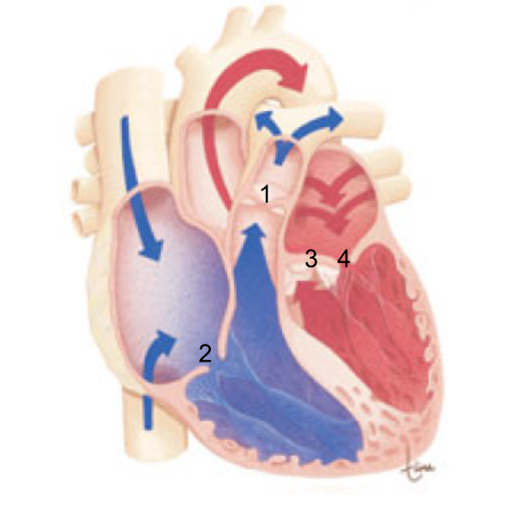
1
2
3
4
The Correct Answer is B
Heart murmurs often originate from abnormal blood flow across heart valves. To understand murmurs, parents must first understand where each valve is located and what it does. The tricuspid valve is one of the atrioventricular (AV) valves, and its location is key in explaining right-sided heart conditions.
Rationale for correct answer
B. The tricuspid valve is located between the right atrium and the right ventricle. It allows blood to flow from the right atrium into the right ventricle and prevents backflow during ventricular contraction. Murmurs related to the tricuspid valve are typically heard along the left lower sternal border.
Rationale for incorrect answers
A. The pulmonic valve is located between the right ventricle and pulmonary artery. It is a semilunar valve, not an atrioventricular valve.
C. The aortic valve is located between the left ventricle and the aorta. It is also a semilunar valve, associated with systemic outflow.
D. The mitral valve is located between the left atrium and left ventricle. This is the left-sided AV valve, not the tricuspid valve.
Test-taking strategy
- First identify whether the valve is atrioventricular (AV) or semilunar.
- Remember:
- Right side AV valve = Tricuspid
- Left side AV valve = Mitral
- Outflow valves = Aortic and Pulmonic
Take-home points
- The tricuspid valve lies between the right atrium and right ventricle.
- AV valves (tricuspid and mitral) are common sources of murmurs due to regurgitation or stenosis.
- Understanding valve location helps parents grasp the cause and significance of congenital murmurs.
Nursing Test Bank
Naxlex Comprehensive Predictor Exams
Related Questions
Correct Answer is C
Explanation
Heart sounds are produced by valve closure and changes in blood flow within the heart. Correct interpretation of heart sounds requires understanding the cardiac cycle, specifically the relationship between atrial contraction, ventricular contraction, and valve movement.
Rationale for correct answer:
C. S1 is directly caused by AV valve closure. It signifies the beginning of ventricular systole. This sound is best heard at the apex of the heart, where the mitral valve is located. Clinically, S1 coincides with the carotid pulse, ventricular contraction, and rising ventricular pressure.
Rationale for incorrect answers:
A. Late diastole is characterized by atrial contraction and ventricular filling. The AV valves are still open at this time, so no S1 occurs yet.
B. Early diastole begins after ventricular relaxation, when the semilunar valves close (aortic and pulmonic valves), producing S2, not SA.
D. Closure of the aortic and pulmonic (semilunar) valves generates the second heart sound (S2), which marks the end of systole and beginning of diastole.
Test-taking strategy:
- Always match valve type to heart sound:
- AV valves closure (mitral, tricuspid) causes SA.
- Semilunar valves closure (aortic, pulmonic) causes SB.
- Use timing clues:
- Systole begins with S1
- Diastole begins with S2
Take home points
- S1 (“lub”) occurs with closure of the mitral and tricuspid valves and marks the beginning of ventricular systole.
- S1 is best heard at the apex and coincides with the carotid pulse.
- S2 (“dub”) results from semilunar valves closure and marks the start of diastole.
- Understanding heart sounds requires linking valve movement, pressure changes, and the cardiac cycle.
Correct Answer is D
Explanation
Physiological splitting of S2 occurs when the aortic (A2) and pulmonic (P2) components of the second heart sound are heard as two distinct sounds during inspiration. This is a normal finding in healthy children and adults, caused by increased venous return during inspiration, which delays closure of the pulmonic valve.
Rationale for correct answer:
D. Document the findings as a normal finding: Physiological splitting is most prominent during deep inspiration and is considered normal, especially in children, adolescents, and young adults. No intervention or provider notification is required unless other abnormal findings are present such as cyanosis, murmurs, or signs of heart failure. Accurate documentation ensures continuity of care and reflects the child’s normal cardiac physiology.
Rationale for incorrect answers:
A. Notify the provider of suspected atrial-septal defect (ASD): ASD can cause fixed splitting of S2, which is present in both inspiration and expiration. Physiological splitting is variable with respiration and does not indicate a septal defect.
B. Notify the provider of suspected pulmonary stenosis: Pulmonary stenosis may produce a loud systolic ejection murmur with fixed splitting, not normal physiological splitting. The described finding does not suggest pathology.
C. Follow institutional policy for initiating an emergency response: This is unnecessary because physiological splitting of S2 is a normal, nonemergent finding.
Test-taking strategy:
- Distinguish physiological vs. pathological splitting:
- Physiological splitting: occurs only with inspiration, varies with respiration, normal in children.
- Pathological splitting: fixed or paradoxical, may indicate ASD, bundle branch block, or pulmonary stenosis.
- If the split changes with breathing and no other abnormalities are present, it is normal.
Take home points
- Physiological splitting of S2 is a normal variation in children and adults during deep inspiration.
- Documenting normal findings is appropriate; no emergency or provider notification is required.
- Fixed or paradoxical splitting, loud murmurs, or other abnormal signs should prompt further evaluation.
- Understanding normal cardiac sounds prevents unnecessary interventions and anxiety.
Whether you are a student looking to ace your exams or a practicing nurse seeking to enhance your expertise , our nursing education contents will empower you with the confidence and competence to make a difference in the lives of patients and become a respected leader in the healthcare field.
Visit Naxlex, invest in your future and unlock endless possibilities with our unparalleled nursing education contents today