Identify the letter of the heart component that is being described in the statement.
Shallow depression that is a remnant of the foramen ovale.
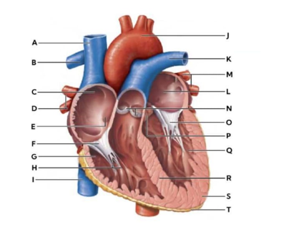
N
E
O
J
The Correct Answer is B
N: Pulmonary valve- The pulmonary valve is a semilunar valve between the right ventricle and pulmonary artery. It prevents backflow into the ventricle during diastole, facilitating blood flow toward the lungs for oxygenation.
E: Fossa ovalis - The fossa ovalis is a shallow, thumb-sized depression located in the interatrial septum (the wall separating the right and left atria). In a developing fetus, the foramen ovale is an open "tunnel" that allows blood to bypass the lungs by flowing directly from the right atrium to the left atrium. Once a baby takes their first breath, the pressure changes in the heart cause a flap of tissue to close over this opening. Over time, it fuses shut, leaving behind the shallow indensee fossa ovalis.
O: chordae tendinae: Chordae tendineae are fibrous cords connecting the atrioventricular valve leaflets (mitral and tricuspid) to papillary muscles. They prevent valve prolapse during ventricular contraction, ensuring unidirectional blood flow.
J: Aortic arch- The aortic arch is the curved portion of the aorta that distributes oxygenated blood from the left ventricle to systemic arteries. It contains baroreceptors and helps regulate blood pressure.
Nursing Test Bank
Naxlex Comprehensive Predictor Exams
Related Questions
Correct Answer is A
Explanation
A. Glucagon: Glucagon acts as a positive inotropic agent by increasing intracellular cyclic AMP (cAMP) in cardiac myocytes, which enhances calcium availability for actin-myosin cross-bridge formation. This results in stronger myocardial contractions and improved cardiac output. It is particularly used in cases of beta-blocker overdose to support cardiac contractility when adrenergic stimulation is blocked.
B. Calcium channel blockers: Calcium channel blockers, such as verapamil and diltiazem, inhibit calcium influx into cardiac and vascular smooth muscle cells. This action reduces myocardial contractility (negative inotropic effect) and slows conduction through the atrioventricular node, which can decrease cardiac output in some patients.
C. Potassium: Potassium primarily influences the resting membrane potential and excitability of cardiac cells. Hyperkalemia can depress myocardial contractility, whereas hypokalemia can predispose to arrhythmias. Potassium itself does not act as a positive inotropic agent.
D. Beta blockers: Beta-adrenergic blockers decrease sympathetic stimulation of the heart by antagonizing beta-1 receptors. This leads to reduced heart rate, decreased myocardial contractility (negative inotropic effect), and lower oxygen demand, making them useful for hypertension and heart failure management but not for increasing contractile force.
Correct Answer is C
Explanation
A. Contractile force increases to compensate for the reduced cardiac output: In early compensatory phases, the heart may attempt to increase contractility via sympathetic stimulation, but in true heart failure, the myocardium is unable to generate sufficient force due to structural or functional impairment.
B. Contractile force increases, leading to an increased end systolic volume: Increased contractility would reduce, not increase, end-systolic volume because more blood is ejected per beat. In heart failure, contractile weakness leads to higher end-systolic volumes, reflecting incomplete emptying of the ventricles.
C. Contractile force is diminished due to damaged cardiomyocytes or cardiomyopathies: Heart failure results from conditions such as myocardial infarction, chronic hypertension, or dilated cardiomyopathy that impair cardiomyocyte function. This reduces the strength of ventricular contraction, decreasing stroke volume and overall cardiac output.
D. Contractile force is not affected in heart failure: Contractile force is significantly affected in heart failure. The weakened myocardium cannot generate sufficient pressure to maintain normal stroke volume, making this statement inaccurate.
E. Contractile force remains the same, but the heart becomes larger: While ventricular dilation can occur in chronic heart failure as a compensatory mechanism (eccentric hypertrophy), the contractile force per myocyte is reduced. Increased chamber size alone does not preserve effective contraction.
Whether you are a student looking to ace your exams or a practicing nurse seeking to enhance your expertise , our nursing education contents will empower you with the confidence and competence to make a difference in the lives of patients and become a respected leader in the healthcare field.
Visit Naxlex, invest in your future and unlock endless possibilities with our unparalleled nursing education contents today