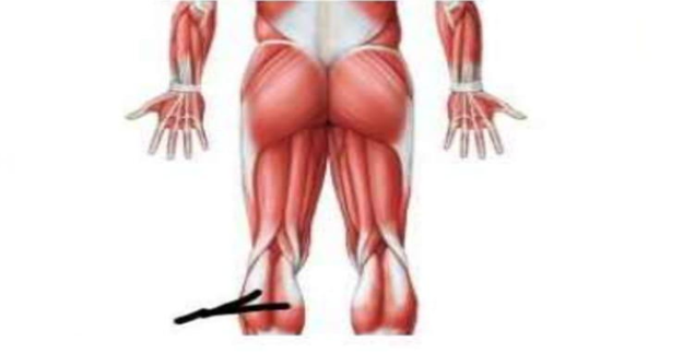
Identify the muscle that is indicated by the leader line in the image below.
Gastrocnemius
Tibialis anterior
Soleus
Fibularis (peroneus) longus
The Correct Answer is A
The marked structure is the gastrocnemius muscle, the most superficial muscle of the posterior compartment of the leg, forming the prominent bulge of the calf. It has two heads (medial and lateral) originating from the femoral condyles and inserting via the Achilles (calcaneal) tendon into the calcaneus. The gastrocnemius crosses both the knee and ankle joints, making it a powerful plantar flexor of the foot and a weak knee flexor. It plays a major role in walking, running, and jumping by providing propulsion during the push-off phase of gait.
A. Gastrocnemius: The gastrocnemius is a large, superficial posterior leg muscle forming the bulk of the calf. It originates from the medial and lateral femoral condyles and joins the soleus to form the Achilles tendon inserting into the calcaneus. Its primary function is plantar flexion of the ankle and assisting knee flexion. Because it is the most prominent posterior calf muscle visible externally, it corresponds to the marked structure.
B. Tibialis anterior: The tibialis anterior is located in the anterior compartment of the leg along the shin. It is responsible for dorsiflexion and inversion of the foot, allowing toe clearance during walking. It lies deep to the anterior skin surface of the tibia and does not form the calf bulge. Compared to the gastrocnemius, it is anterior and functions opposite in ankle movement.
C. Soleus: The soleus is a deep posterior leg muscle located beneath the gastrocnemius. It originates from the tibia and fibula and also inserts into the calcaneus via the Achilles tendon. It is a powerful plantar flexor, especially during standing and postural control. However, it is not as superficially visible as the gastrocnemius and does not form the prominent calf contour seen in the marked structure.
D. Fibularis (peroneus) longus: The fibularis longus is located in the lateral compartment of the leg. It originates from the fibula and functions in eversion and plantar flexion of the foot. It runs along the lateral aspect of the leg and foot, contributing to arch support. Unlike the gastrocnemius, it does not form the posterior calf bulge and is more lateral and deep in position.
Nursing Test Bank
Naxlex Comprehensive Predictor Exams
Related Questions
Correct Answer is A
Explanation
The marked structure is the masseter muscle, one of the primary muscles of mastication located on the lateral aspect of the mandible. It originates from the zygomatic arch and inserts onto the lateral surface of the ramus and angle of the mandible. The masseter is one of the strongest muscles in the body relative to its size and is essential for forceful elevation of the mandible during chewing. It works in coordination with the temporalis and pterygoid muscles to produce efficient grinding and crushing of food during mastication.
A. Masseter: The masseter is a thick, rectangular muscle situated over the lateral surface of the mandibular ramus. It elevates the mandible, producing powerful jaw closure required for chewing tough food. It has superficial and deep layers and is easily visible when the jaw is clenched. Its location over the angle of the jaw and strong vertical fibers make it the correct structure.
B. Zygomatic: The zygomatic region refers to the zygomatic bone (cheekbone), which forms the prominence of the cheek and part of the orbital rim. It is a bone, not a muscle, and serves as an attachment site for facial muscles. Unlike the masseter, it does not contract or contribute to jaw movement.
C. Buccinator: The buccinator is a thin, flat muscle located in the cheek. It assists in compressing the cheek against the teeth, aiding in chewing by keeping food between the occlusal surfaces. It is also involved in blowing and whistling. Unlike the masseter, it is deep and does not produce strong jaw elevation.
D. Temporalis: The temporalis is a fan-shaped muscle located on the lateral skull in the temporal fossa. It elevates and retracts the mandible and plays a key role in closing the jaw. Although it is also a muscle of mastication, it is positioned superiorly on the skull rather than over the lateral jaw like the masseter.
Correct Answer is B
Explanation
The nervous system is a highly specialized communication network responsible for detecting internal and external stimuli, processing information, and coordinating appropriate responses. It is composed of the central nervous system (brain and spinal cord) and peripheral nervous system (nerves and ganglia). Its primary mode of communication is rapid electrical signaling via neurons, which allows precise control of bodily functions. This system works closely with the endocrine system to maintain homeostasis.
A. Contracts and generates a force to cause bodily motion: contraction and force generation are functions of the muscular system, not the nervous system. Skeletal muscles contract in response to neural stimulation, but they are the effectors rather than the controllers. The nervous system only initiates and regulates these contractions. This describes muscle function rather than neural function.
B. Controls and coordinates body activities by transmitting electrical signals: the nervous system regulates body functions through rapid transmission of electrical impulses along neurons. These signals allow communication between different parts of the body and enable coordination of movement, sensation, and autonomic processes. The brain and spinal cord integrate information and generate appropriate responses. This makes the nervous system the primary control and coordination system of the body.
C. Produces hormones that directly contract skeletal muscle: hormone production is primarily the role of the endocrine system, not the nervous system. While some endocrine glands are regulated by the nervous system (e.g., hypothalamus and pituitary), hormones themselves do not directly cause skeletal muscle contraction. Muscle contraction is triggered by motor neuron stimulation via neurotransmitters, not hormones.
D. Stores nutrients for energy use during muscle contraction: nutrient storage is primarily the function of tissues such as the liver, adipose tissue, and skeletal muscle. Glycogen and fat serve as energy reserves, but this is not a function of the nervous system. The nervous system may regulate metabolic activity but does not store energy substrates. This option describes metabolic storage functions rather than neural activity.
Whether you are a student looking to ace your exams or a practicing nurse seeking to enhance your expertise , our nursing education contents will empower you with the confidence and competence to make a difference in the lives of patients and become a respected leader in the healthcare field.
Visit Naxlex, invest in your future and unlock endless possibilities with our unparalleled nursing education contents today