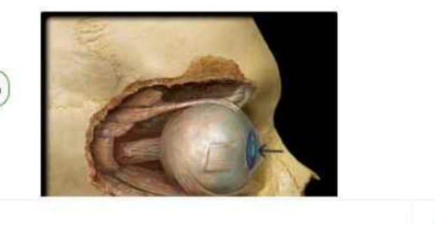
Identify the part of the eye that is highlighted greenish blue and indicated by the arrow in the image below (it is the small circle, that can constrict and dilate).
Cornea
Pupil
Lens
Iris
The Correct Answer is B
The marked structure is the pupil, the central circular opening within the iris through which light enters the eye. Although commonly perceived as a black structure, the pupil is actually an aperture rather than a physical tissue. Its diameter changes continuously in response to light intensity and autonomic nervous system stimulation through the actions of the sphincter pupillae and dilator pupillae muscles located within the iris. Regulation of pupil size is essential for controlling the amount of light reaching the retina and optimizing visual acuity under varying environmental conditions.
A. Cornea: The cornea is the transparent, avascular anterior portion of the fibrous tunic of the eye that covers the iris, pupil, and anterior chamber. It provides approximately two-thirds of the eye’s refractive power by bending incoming light toward the retina. Unlike the pupil, the cornea is a physical structure composed of specialized layers of tissue and does not constrict or dilate in response to light.
B. Pupil: The pupil is the circular opening located at the center of the iris and serves as the gateway through which light enters the eye. Its size changes through pupillary constriction (miosis) and dilation (mydriasis), allowing regulation of retinal light exposure. Parasympathetic stimulation causes constriction, whereas sympathetic stimulation causes dilation. Because the marked structure is the small circular opening that changes diameter in response to light, it is the pupil.
C. Lens: The lens is a transparent, biconvex structure located directly posterior to the iris and pupil. It functions by altering its shape during accommodation to focus light rays precisely onto the retina for near and distant vision. Unlike the pupil, the lens is a solid anatomical structure and does not change size to regulate light entry. Its role is optical focusing rather than light regulation.
D. Iris: The iris is the pigmented, circular structure surrounding the pupil and responsible for determining eye color. It contains smooth muscle fibers arranged as the sphincter pupillae and dilator pupillae muscles, which control pupil diameter. While the iris performs the mechanical action that changes pupil size, the opening that actually constricts and dilates is the pupil itself. Therefore, the iris surrounds the marked structure but is not the structure being identified.
Nursing Test Bank
Naxlex Comprehensive Predictor Exams
Related Questions
Correct Answer is A
Explanation
The marked structure is located within smooth muscle tissue, which is composed of spindle-shaped, non-striated muscle cells found in the walls of hollow organs such as blood vessels, intestines, and the urinary bladder. These cells are responsible for involuntary movements regulated by the autonomic nervous system. In histological sections, smooth muscle is identified by elongated, spindle-shaped cells with centrally placed, cigar-shaped nuclei and absence of striations. The arrow in this image is pointing to the elongated central nucleus of a smooth muscle cell.
A. Smooth muscle cell: The image shows elongated, spindle-shaped cells arranged in parallel bundles with centrally located, elongated nuclei. These features are characteristic of smooth muscle tissue, which lacks striations due to the non-organized arrangement of actin and myosin filaments. The nucleus is the most prominent visible structure in histological sections, appearing as a dark, cigar-shaped structure in the center of each cell. Smooth muscle is responsible for involuntary contraction in organs such as blood vessels and the gastrointestinal tract, controlling processes like peristalsis and vasoconstriction.
B. Skeletal muscle cell: Skeletal muscle fibers are long, cylindrical, and multinucleated with nuclei located peripherally rather than centrally. They also display prominent cross-striations due to organized sarcomeres. Unlike the tissue shown, skeletal muscle is voluntary and found attached to bones for movement. The absence of striations and peripheral nuclei rules out skeletal muscle in this image.
C. Cardiac muscle cell: Cardiac muscle cells are branched, striated, and contain a single centrally located nucleus. They also show intercalated discs that connect adjacent cells. While they have central nuclei similar to smooth muscle, the presence of striations and branching distinguishes them. The tissue in the image lacks these features, making cardiac muscle incorrect.
D. Connective tissue fibroblast: Fibroblasts are spindle-shaped cells found in connective tissue and may appear elongated under the microscope. However, they are typically embedded within a collagen-rich extracellular matrix rather than tightly packed parallel muscle fibers. Unlike smooth muscle cells, they do not form organized contractile bundles or show uniform alignment as seen in this image.
Correct Answer is A
Explanation
The marked structure is the scapula, a flat, triangular bone located on the posterior aspect of the thoracic cage, commonly referred to as the shoulder blade. It lies over ribs 2–7 and forms the posterior component of the shoulder girdle. The scapula plays a central role in upper limb mobility by serving as an attachment site for multiple muscles that control shoulder movement and stabilization. It articulates with the clavicle at the acromioclavicular joint and with the humerus at the glenohumeral (shoulder) joint.
A. Scapula: The scapula is a flat, triangular bone positioned on the posterior thoracic wall. It contains important anatomical landmarks such as the spine, acromion, coracoid process, and glenoid cavity, which participate in shoulder articulation and muscle attachment. It allows a wide range of shoulder movements including elevation, rotation, and abduction through coordinated muscular action. Since the marked structure lies on the posterior upper back forming the shoulder blade, it corresponds to the scapula.
B. Clavicle: The clavicle is a long, S-shaped bone located anteriorly at the base of the neck. It connects the sternum to the scapula, acting as a strut that stabilizes the shoulder girdle. Its main function is to maintain shoulder position and allow upper limb mobility away from the trunk. Unlike the scapula, it is a horizontal anterior bone rather than a flat posterior structure.
C. Humerus: The humerus is the long bone of the upper arm extending from the shoulder to the elbow joint. It serves as the main structural bone for arm movement and muscle attachment. It articulates with the scapula at the glenoid cavity to form the shoulder joint. However, it is located in the arm rather than forming the posterior shoulder blade itself.
D. Ribs: The ribs are curved, flat bones forming the thoracic cage that protects the heart and lungs. They articulate posteriorly with the thoracic vertebrae and anteriorly with the sternum (via costal cartilage). Their primary function is protection and respiratory movement. Unlike the scapula, they are part of the thoracic cage rather than the shoulder girdle.
Whether you are a student looking to ace your exams or a practicing nurse seeking to enhance your expertise , our nursing education contents will empower you with the confidence and competence to make a difference in the lives of patients and become a respected leader in the healthcare field.
Visit Naxlex, invest in your future and unlock endless possibilities with our unparalleled nursing education contents today