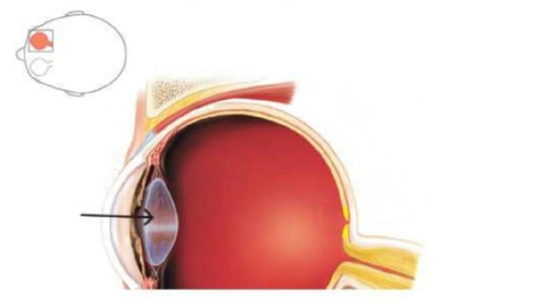
Identify the part of the eye indicated by the arrow in the image below.
Cornea
Retina
Iris
Lens
The Correct Answer is D
The marked structure is the lens, a transparent, biconvex, avascular structure located posterior to the iris and anterior to the vitreous body. It is suspended by zonular fibers (suspensory ligaments) attached to the ciliary body. The lens plays a critical role in vision by providing fine focusing of light onto the retina through the process of accommodation. Its curvature changes depending on whether the eye is focusing on near or distant objects, allowing precise image formation.
A. Cornea: The cornea is the transparent, dome-shaped anterior surface of the eye that provides most of the eye’s refractive power. It is the first structure that light passes through and contributes significantly to bending light toward the retina. Unlike the lens, it is external, fixed in shape, and continuous with the sclera, serving both protective and optical roles.
B. Retina: The retina is the innermost neural layer lining the posterior aspect of the eye. It contains photoreceptor cells (rods and cones) that convert light into electrical signals sent via the optic nerve to the brain. Unlike the lens, it does not focus light but instead processes visual information after image formation occurs.
C. Iris: The iris is the pigmented muscular structure located anterior to the lens and posterior to the cornea. It regulates pupil size to control the amount of light entering the eye through contraction and relaxation of its smooth muscles. Unlike the lens, it does not contribute to focusing light but only regulates light entry.
D. Lens: The lens is a transparent, flexible, biconvex structure located directly behind the iris. It fine-tunes the focusing of light rays onto the retina through accommodation, changing its curvature via the ciliary muscles. It plays a key role in sharp image formation at varying distances. Its central posterior position relative to the iris makes it the correct structure.
Nursing Test Bank
Naxlex Comprehensive Predictor Exams
Related Questions
Correct Answer is C
Explanation
The human skeleton is divided into two major parts: the axial skeleton and the appendicular skeleton. The axial skeleton forms the central axis of the body and is responsible for supporting the head, neck, and trunk. It also provides protection for vital organs such as the brain, spinal cord, and thoracic organs. Understanding this division is essential for identifying bone groups and their functional roles in movement, support, and protection.
A. Radius, ulna, carpals, and phalanges: These bones are part of the upper limb and therefore belong to the appendicular skeleton. The radius and ulna form the forearm, while the carpals and phalanges make up the wrist and fingers. Their primary function is to facilitate movement and manipulation of objects. Since they are located in the limbs rather than the central body axis, they are not part of the axial skeleton.
B. Femur, tibia, fibula, and patella: These bones belong to the lower limb and are part of the appendicular skeleton. The femur is the thigh bone, the tibia and fibula form the lower leg, and the patella is the kneecap. Together, they support weight-bearing and locomotion. However, they are not part of the central axis of the body, so they are excluded from the axial skeleton.
C. Skull, hyoid bone, thoracic cage, and vertebral column: these structures form the axial skeleton. The skull protects the brain, the vertebral column houses the spinal cord, the thoracic cage (ribs and sternum) protects the heart and lungs, and the hyoid bone supports tongue and swallowing functions. Collectively, these structures form the central framework of the body and provide protection and support for vital organs.
D. Scapula, clavicle, humerus, and pelvic bones: These bones are part of the appendicular skeleton, which includes the girdles and limbs. The scapula and clavicle form the shoulder girdle, the humerus is the upper arm bone, and the pelvic bones support the lower trunk and connect the lower limbs to the axial skeleton. Their primary role is movement and attachment of limbs rather than central body support, so they are not part of the axial skeleton.
Correct Answer is C
Explanation
Neuronal excitability depends on changes in membrane potential, which is the electrical difference across the cell membrane. At rest, neurons maintain a negative resting membrane potential due to ion gradients established by the sodium-potassium pump and selective membrane permeability. Depolarization is the process by which the membrane potential becomes less negative and moves toward zero or positive values. This event is essential for initiating action potentials and allowing nerve impulse transmission along neurons.
A. Na⁺ channels close and Na⁺ ions cannot enter the cell: closure of sodium channels would prevent sodium influx and therefore maintain or reinforce the resting membrane potential. Without sodium entry, the inside of the neuron remains negatively charged relative to the outside. Depolarization specifically requires an influx of positive ions, not their exclusion. This describes inhibition of depolarization rather than its initiation.
B. K⁺ channels open and K⁺ diffuses into the cell: potassium movement typically involves efflux, not influx, during neuronal activity. When potassium channels open, K⁺ generally leaves the cell, contributing to repolarization or hyperpolarization rather than depolarization. The movement of positive ions out of the cell increases negativity inside the membrane.
C. Na⁺ channels open and Na⁺ ions diffuse into the cell: depolarization occurs when voltage-gated sodium channels open and allow Na⁺ ions to flow into the neuron. Sodium ions enter the cell down their electrochemical gradient, making the inside of the membrane less negative. This rapid influx of positive charge initiates the rising phase of the action potential. It is the fundamental event that triggers neuronal firing.
D. Chloride ions enter the cell causing hyperpolarization: chloride influx typically makes the inside of the neuron more negative, leading to hyperpolarization rather than depolarization. Increased negativity moves the membrane potential further from the threshold required for action potential generation. Chloride entry stabilizes or inhibits neuronal firing.
Whether you are a student looking to ace your exams or a practicing nurse seeking to enhance your expertise , our nursing education contents will empower you with the confidence and competence to make a difference in the lives of patients and become a respected leader in the healthcare field.
Visit Naxlex, invest in your future and unlock endless possibilities with our unparalleled nursing education contents today