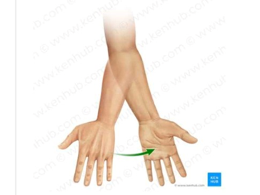
Looking at the arrow, the patient is moving his hand from which position to the following position?
Pronation to supination
Supination to pronation
Pronation
Supination
The Correct Answer is A
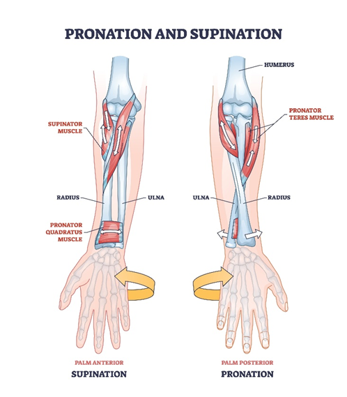
Forearm rotation involves the proximal radioulnar joint where the radius crosses over the ulna. The movement of the palm from a downward-facing (posterior) position to an upward-facing (anterior) position is supination, while the reverse is pronation. These actions are essential for manual dexterity and are facilitated by the biceps brachii and supinator muscles.
A. Pronation to supination: The image shows the hand starting with the dorsum visible and the palm facing down, which is the pronated position. The green arrow indicates a lateral rotation that brings the palm to face upward. This transition is the anatomical definition of moving from a state of pronation to supination.
B. Supination to pronation: This movement would involve starting with the palm facing up and rotating the thumb medially until the palm faces down. The arrow in the provided image moves in the opposite direction. Therefore, this description does not match the visual evidence of the hand's trajectory.
C. Pronation: This term refers to the static state of having the palm face downward or the specific act of turning it down. While the hand begins in this position, the arrow indicates a dynamic change toward a different orientation. It does not account for the completed movement shown in the second hand position.
D. Supination: This refers to the final position shown where the palm is facing anteriorly, often described as the anatomical position for the forearm. However, the question asks for the transition represented by the arrow. "Supination" alone describes a state rather than the "from-to" action depicted.
Nursing Test Bank
Naxlex Comprehensive Predictor Exams
Related Questions
Correct Answer is A
Explanation
Auscultation of bowel sounds requires the use of the diaphragm because peristaltic noises are predominantly high-pitched. The nurse must apply only light pressure to avoid stimulating the underlying smooth muscle, which could artificially increase motility. Systematic assessment begins in the right lower quadrant near the ileocecal valve, where sounds are typically most audible.
A. Hold the diaphragm of the stethoscope lightly against the abdomen in each quadrant: The diaphragm is the correct tool for capturing high-frequency bowel sounds. Light contact ensures the nurse hears the patient's baseline gastrointestinal activity without causing discomfort or reactive peristalsis. This is the standard, evidence-based technique for an abdominal assessment.
B. Hold the bell of the stethoscope lightly against the abdomen in each quadrant: The bell of the stethoscope is designed to pick up low-pitched sounds, such as vascular bruits or heart murmurs. It is not the appropriate instrument for hearing the clicks and gurgles of the intestines. Using the bell would lead to an incomplete or muffled assessment.
C. Press the diaphragm of the stethoscope firmly against the abdomen in each quadrant: Firm pressure can cause the patient to guard their muscles and may physically stimulate the bowel, producing sounds that were not present at rest. It can also cause pain if the patient has underlying tenderness. Light pressure is preferred to maintain a neutral diagnostic environment.
D. Press the bell of the stethoscope firmly against the abdomen in each quadrant: Pressing the bell firmly against the skin actually converts it into a diaphragm, but it remains ineffective for the high-pitched sounds of the gut. Furthermore, the firm pressure violates the principle of not stimulating the abdomen before completing the auscultation.
Correct Answer is ["A","B","C","D"]
Explanation
The neurocranium consists of 8 bones that form the protective vault surrounding the brain. It is distinguished from the viscerocranium, which comprises the facial skeleton. These bones are joined by sutures, which are immobile fibrous joints, providing structural integrity to the skull.
A. Occipital: This bone forms the posterior and inferior base of the cranium and contains the foramen magnum. It articulates with the atlas of the vertebral column. It is a primary component of the cranial vault protecting the cerebellum.
B. Temporal: These paired bones form the lateral walls and base of the skull, housing the structures of the inner ear. They articulate with the mandible at the temporomandibular joint. They are essential components of the lateral neurocranium.
C. Frontal: This bone forms the forehead and the superior portion of the orbit and the anterior cranial fossa. It contains the frontal sinuses and provides protection for the frontal lobes. It is a major constituent of the cranium.
D. Parietal: These paired bones form the bulk of the superior and lateral vault of the skull. They meet at the sagittal suture and articulate with the frontal and occipital bones. They are fundamental parts of the cranial structure.
E. Zygomatic: Known as the cheekbones, these are components of the viscerocranium or facial skeleton rather than the neurocranium. They form the lateral wall and floor of the orbit. They do not contribute to the protective brain case.
Whether you are a student looking to ace your exams or a practicing nurse seeking to enhance your expertise , our nursing education contents will empower you with the confidence and competence to make a difference in the lives of patients and become a respected leader in the healthcare field.
Visit Naxlex, invest in your future and unlock endless possibilities with our unparalleled nursing education contents today