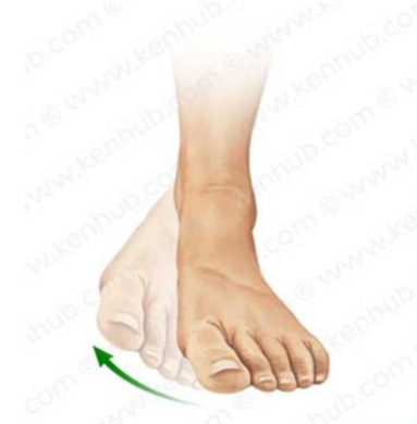
Looking at the arrow, what is the direction of the foot in the picture.
Eversion
Pronation
Supination
Inversion
The Correct Answer is D
Inversion is a complex movement of the foot that tilts the sole medially toward the midline of the body. This action primarily occurs at the subtalar and midtarsal joints, facilitated by the tibialis anterior and posterior muscles. It is an essential component of the musculoskeletal exam to evaluate ligamentous stability and neuromuscular control of the ankle.
A. Eversion: Eversion involves tilting the sole of the foot laterally, away from the body's midline. This movement is the anatomical opposite of the inward tilt shown in the picture. It primarily assesses the strength of the peroneal muscles and the integrity of the lateral ankle stabilizers.
B. Pronation: In the context of the foot, pronation is a triplanar movement involving eversion, abduction, and dorsiflexion. It results in a flattening of the medial longitudinal arch. The image focuses on a simple medial tilt, which is a component of supination rather than pronation.
C. Supination: While inversion is a major component of foot supination, supination also includes adduction and plantarflexion. In clinical terminology, when the sole specifically turns inward as shown by the arrow, the most precise term for that specific directional movement is inversion.
D. Inversion: The image depicts the foot being turned so that the plantar surface faces toward the other foot. This specific medial rotation of the foot at the ankle is defined as inversion. This maneuver is frequently tested to check for sprain-related tenderness in the lateral ligaments.
Nursing Test Bank
Naxlex Comprehensive Predictor Exams
Related Questions
Correct Answer is D
Explanation
Abdominal examination requires maximal relaxation of the rectus abdominis muscles to allow for accurate palpation of deep structures. Placing a small pillow under the head and having the patient flex the knees reduces tension on the abdominal wall. This position prevents voluntary guarding, which can otherwise obscure underlying masses or organomegaly.
A. Ask the patient to refrain from voiding before the exam: An overdistended bladder can cause significant discomfort during palpation and may be mistaken for an abdominal mass or suprapubic tenderness. Patients should be encouraged to empty their bladder immediately prior to the assessment to ensure comfort and diagnostic accuracy.
B. Position the patient's arms above the head: Placing arms above the head stretches the abdominal musculature, increasing wall tension and making deep palpation more difficult and uncomfortable. The arms should remain at the patient's sides or folded across the chest to promote the most relaxed state possible.
C. Examine painful areas on the abdomen first: Assessing tender areas at the start of the exam causes the patient to tense their muscles in anticipation of pain, making the rest of the assessment unreliable. Standard protocol dictates that the nurse should examine painful quadrants last to maintain muscle relaxation and patient trust.
D. Ask the patient to bend their knees slightly: Flexing the knees and hips relaxes the tension in the abdominal wall muscles. This mechanical shift makes the abdomen softer and more accessible for the clinician to perform light and deep palpation without resistance. It is a fundamental step in abdominal examination preparation.
Correct Answer is A
Explanation
The tympanic membrane is a thin, semitransparent partition separating the external auditory canal from the middle ear. During otoscopy, a healthy drum exhibits a pearly grey or translucent appearance with a distinct cone of light reflecting anteroinferiorly. Its concave morphology is maintained by the attachment of the malleus handle at the umbo.
A. Concave and pearly grey: This is the classic clinical description of a healthy eardrum. The concave shape results from the pull of the auditory ossicles, and the pearly grey color indicates the absence of middle ear effusion or infection. It reflects a normal, air-filled middle ear cavity.
B. Opaque and red: Redness and opacity suggest acute otitis media, where the membrane becomes hyperemic and thickened due to inflammation. This finding is pathological and often associated with pain and fever. It indicates a loss of transparency and healthy vascular regulation.
C. Convex and slightly white: A convex or bulging appearance indicates increased pressure within the middle ear, often from purulent fluid or effusion. A white appearance can signify myringosclerosis or scarring. It is not the expected finding for a healthy, functioning membrane.
D. Straight and pink: The tympanic membrane is naturally curved rather than straight. While mild pinkness can sometimes occur with crying or irritation, a truly healthy drum is grey. A straight appearance would suggest a lack of structural tension from the ossicular chain.
Whether you are a student looking to ace your exams or a practicing nurse seeking to enhance your expertise , our nursing education contents will empower you with the confidence and competence to make a difference in the lives of patients and become a respected leader in the healthcare field.
Visit Naxlex, invest in your future and unlock endless possibilities with our unparalleled nursing education contents today