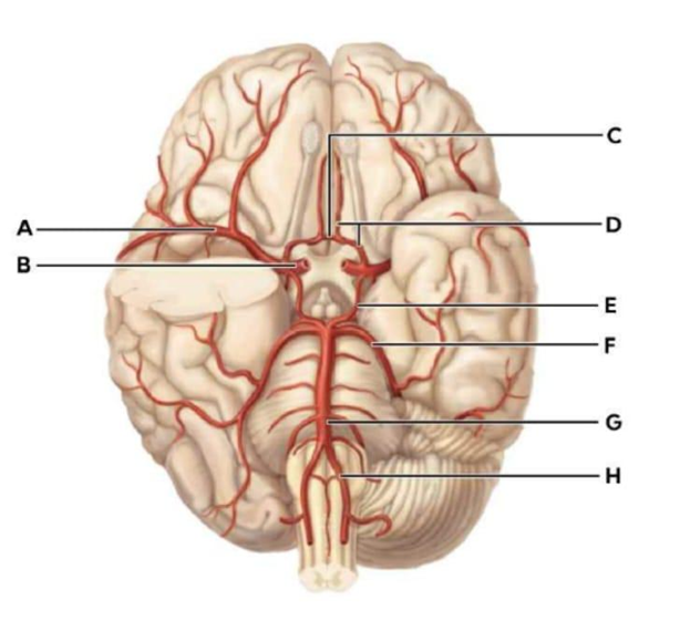
Match the letter to its blood vessel:
| C | dropdown |
| B | dropdown |
| G | dropdown |
| H | dropdown |
The Correct Answer is {"dropdown-group-1":"A","dropdown-group-2":"B","dropdown-group-3":"C","dropdown-group-4":"D"}
C: Anterior communicating artery
B: Internal Carotid Artery
G: Basilar Artery
H: Vertebral artery
A. C (Anterior Communicating Artery): The anterior communicating artery is a short midline vessel that connects the right and left anterior cerebral arteries. It forms the anterior portion of the Circle of Willis. Its main role is to allow blood to cross from one side of the cerebral circulation to the other, providing collateral flow if one internal carotid artery becomes narrowed or blocked.
B. B (Internal Carotid Artery): The internal carotid arteries are major contributors to the Circle of Willis. After entering the cranial cavity through the carotid canal, each internal carotid artery gives rise to: The anterior cerebral artery, the middle cerebral artery and the posterior communicating artery. Within the Circle of Willis, the internal carotid arteries form the lateral portions of the circle and contribute to the anterior circulation of the brain.
C. G (Basilar Artery): The basilar artery is formed by the union of the right and left vertebral arteries at the pontomedullary junction. It ascends along the ventral surface of the pons and terminates by dividing into the right and left posterior cerebral arteries. These posterior cerebral arteries contribute to the posterior portion of the Circle of Willis, connecting to the internal carotid system via the posterior communicating arteries.
D. H (Vertebral Artery): Each vertebral artery arises from the subclavian artery and ascends through the transverse foramina of the cervical vertebrae before entering the cranial cavity via the foramen magnum. The two vertebral arteries unite to form the basilar artery. They supply the basilar artery, which contributes to the posterior cerebral circulation within the circle.
Nursing Test Bank
Naxlex Comprehensive Predictor Exams
Related Questions
Correct Answer is D
Explanation
A. Both venoconstriction and vasoconstriction decrease flow: While vasoconstriction of arterioles reduces blood flow by increasing resistance, venoconstriction does not primarily act to decrease flow. Instead, it mobilizes blood from the venous reservoir to the heart, increasing preload and cardiac output, which can enhance systemic flow.
B. Venoconstriction increases resistance and decreases flow, while vasoconstriction does the opposite: Venoconstriction primarily reduces venous compliance rather than increasing resistance in the arterial system. It shifts blood toward the central circulation, raising venous return, stroke volume, and ultimately cardiac output. Arteriolar vasoconstriction increases resistance and reduces flow to downstream tissues.
C. Both venoconstriction and vasoconstriction increase flow: This is partially true for venoconstriction because it increases venous return and can enhance cardiac output. However, vasoconstriction reduces blood flow in arterioles by increasing resistance, so the effect is opposite in the arterial system.
D. Venoconstriction decreases resistance and increases flow, while vasoconstriction increases resistance and decreases flow: Venoconstriction reduces the capacitance of veins, mobilizing stored blood toward the heart, which increases preload, cardiac output, and systemic blood flow. In contrast, vasoconstriction in arterioles raises resistance and limits blood flow to specific tissues. This reflects the fundamental difference between venous and arterial control of circulation.
Correct Answer is A
Explanation
A. Presence of atherosclerotic plaques: Atherosclerotic plaques narrow and irregularly distort the arterial lumen, disrupting normal laminar blood flow. The uneven surface and reduced vessel diameter increase flow velocity and create eddy currents, leading to turbulence. This turbulent flow is often auscultated as a bruit and contributes to endothelial injury and thrombus formation.
B. Slow and smooth blood flow: Slow, steady flow typically promotes laminar movement of blood layers with minimal mixing or disturbance. Laminar flow is characterized by smooth, parallel layers and occurs in healthy vessels without obstruction. It does not generate turbulence under normal physiologic conditions.
C. Decreased heart workload: A reduced cardiac workload generally lowers cardiac output and flow velocity. Lower velocity flow is less likely to exceed the critical Reynolds number required to produce turbulence, especially in vessels without structural abnormalities.
D. Increased venous compliance: Increased venous compliance allows veins to expand and accommodate more blood at lower pressures. This tends to reduce flow velocity and maintain smoother flow patterns. Turbulence is more commonly associated with high velocity and narrowed arterial segments rather than compliant venous systems.
Whether you are a student looking to ace your exams or a practicing nurse seeking to enhance your expertise , our nursing education contents will empower you with the confidence and competence to make a difference in the lives of patients and become a respected leader in the healthcare field.
Visit Naxlex, invest in your future and unlock endless possibilities with our unparalleled nursing education contents today