Provided is an ECG image of sinus tachycardia.
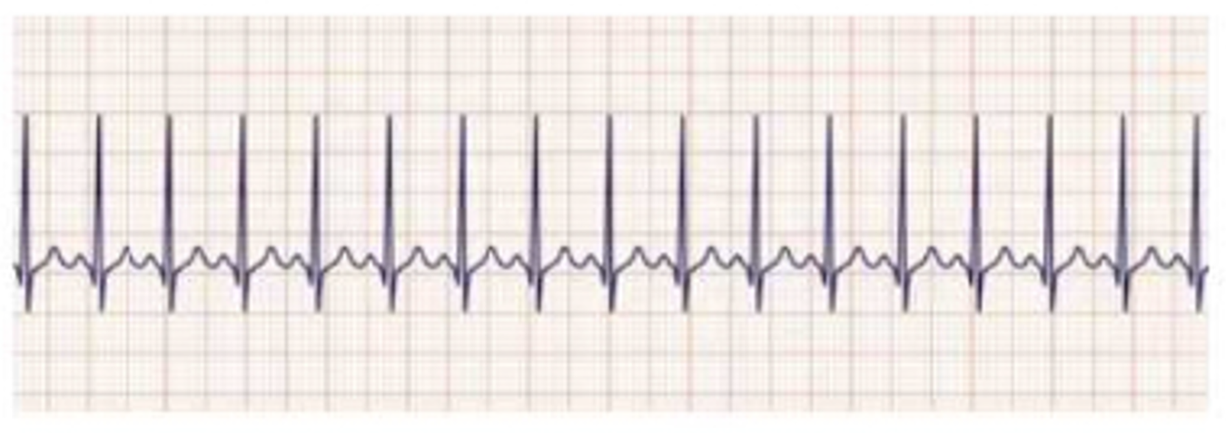
Identify the wave pattern and describe its characteristics.
The wave pattern shows a regular sinus rhythm with a heart rate greater than 100 beats per minute.
The wave pattern shows a regular rhythm with a heart rate between 60 and 100 beats per minute.
The wave pattern shows an irregular rhythm with P waves that change in morphology.
The wave pattern shows a rapid rate with no visible P waves and narrow QRS complexes.
The Correct Answer is A
Choice A rationale
Sinus tachycardia is defined by a heart rate exceeding 100 beats per minute, originating from the sinoatrial node. The rhythm remains regular because the electrical impulses follow the normal conduction pathway through the atria and ventricles. Each QRS complex is preceded by a visible P wave, and the PR interval remains within the normal range of 0.12 to 0.20 seconds. This rhythm is often a physiological response to stress, fever, exercise, or pain.
Choice B rationale
A heart rate between 60 and 100 beats per minute with a regular rhythm defines normal sinus rhythm. In this state, the sinoatrial node fires at a standard physiological pace, and the electrical conduction through the heart is unimpeded. Because sinus tachycardia specifically refers to a rate that is faster than this normal range, this choice does not accurately describe the wave pattern in question. It represents a healthy cardiac electrical state rather than a tachycardic one.
Choice C rationale
An irregular rhythm with changing P wave morphology is characteristic of rhythms like wandering atrial pacemaker or multifocal atrial tachycardia. These conditions involve multiple ectopic foci within the atria competing for dominance, rather than a single impulse from the sinoatrial node. In true sinus tachycardia, the P waves should look identical to one another because they are all generated from the same location. Therefore, this description is inconsistent with the diagnostic criteria for sinus tachycardia.
Choice D rationale
A rapid rate without visible P waves and narrow QRS complexes is typical of supraventricular tachycardia or atrial fibrillation, depending on the regularity of the rhythm. In sinus tachycardia, the P wave must be present and clearly associated with the QRS complex because the rhythm originates from the sinus node. The absence of P waves suggests that the rhythm is originating from a different part of the atrium or the atrioventricular junction, which is incorrect.
Nursing Test Bank
Naxlex Comprehensive Predictor Exams
Related Questions
Correct Answer is D
Explanation
Choice A rationale
Sinus bradycardia is defined by a heart rate below 60 beats per minute with a regular rhythm and discernible P waves. In this case, the client has a heart rate of 92 per minute and an irregular rhythm, which immediately rules out bradycardia. Furthermore, sinus rhythms must have identifiable P waves and measurable PR intervals, both of which are absent in this client's presentation, pointing toward a more chaotic supraventricular origin.
Choice B rationale
First-degree heart block is characterized by a consistent delay in conduction between the atria and ventricles, resulting in a PR interval greater than 0.20 seconds. However, the rhythm remains regular, and P waves must be clearly visible and associated with every QRS complex. The client in the scenario has an irregular rhythm and unidentifiable P waves, which is inconsistent with the stable, albeit delayed, conduction seen in a first-degree block.
Choice C rationale
Supraventricular tachycardia usually manifests as a very rapid, regular rhythm with rates often exceeding 150 beats per minute. While P waves may be difficult to see because they are buried in the preceding T waves, the hallmark is the absolute regularity of the R-to-R intervals. The client's rhythm is described as irregular, which is the primary clinical feature that distinguishes atrial fibrillation from the regular, rapid pacing of a supraventricular tachycardia.
Choice D rationale
Atrial fibrillation is defined by the absence of discrete P waves and an irregularly irregular ventricular rhythm. The fibrillatory waves from the atria do not produce a measurable PR interval because there is no organized atrial depolarization. The QRS duration of 0.10 seconds is within the normal range of 0.06 to 0.12 seconds, indicating that ventricular conduction is still following the normal pathways once the atrioventricular node allows an impulse through.
Correct Answer is B
Explanation
Choice A rationale
Administering nitroglycerin is a standard treatment for chest pain because it induces vasodilation of the coronary arteries. However, prioritizing it over oxygen in a patient with an oxygen saturation of 89 percent is incorrect. Nitroglycerin can significantly lower blood pressure, and it should only be administered after assessing hemodynamic stability. In this clinical scenario, addressing the documented hypoxemia takes precedence to ensure myocardial tissue receives adequate oxygenation to prevent further ischemic damage.
Choice B rationale
Supplemental oxygen is the priority because the patient is hypoxemic, with a saturation below the normal range of 95 to 100 percent. Oxygen therapy increases the partial pressure of oxygen in the blood, enhancing delivery to the myocardium. Correcting hypoxemia reduces the workload on the heart and limits the size of the infarction. Current guidelines emphasize maintaining oxygen saturation at or above 94 percent to optimize cellular respiration and prevent systemic metabolic acidosis.
Choice C rationale
The supine position is not ideal for a patient experiencing chest pain and respiratory distress because it increases venous return and cardiac preload, potentially worsening pulmonary congestion. Positioning the patient in semi-Fowler or high-Fowler position is usually preferred to facilitate lung expansion. While morphine is used for pain that is unresponsive to nitrates, it is not the first priority. Oxygenation must be addressed immediately to mitigate the underlying cause of the myocardial hypoxia.
Choice D rationale
Thrombolytic therapy is a critical intervention for ST-segment elevation myocardial infarction when percutaneous intervention is unavailable, but it is never initiated before establishing vascular access and confirming the diagnosis via a 12-lead ECG. Furthermore, safety screening for contraindications like recent surgery or bleeding disorders is mandatory. Starting this treatment without basic stabilization, such as oxygen administration for a desaturating patient, violates the primary assessment sequence of airway, breathing, and circulation.
Whether you are a student looking to ace your exams or a practicing nurse seeking to enhance your expertise , our nursing education contents will empower you with the confidence and competence to make a difference in the lives of patients and become a respected leader in the healthcare field.
Visit Naxlex, invest in your future and unlock endless possibilities with our unparalleled nursing education contents today