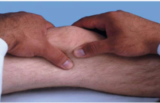
The advanced practice registered nurse (APRN) assesses the knee of a patient who presents with swelling of their right knee. The APRN places their thumb and index finger of their right hand on each side of the patella; with the left hand the APRN compresses the suprapatellar recess against the femur (as shown in the figure below). The APRN is able to palpate ejected fluid or "ballooning" of fluid. The APRN documents a positive "balloon sign", which is associated with:
Bursitis
Minor effusion
Rheumatoid arthritis
Major effusion
The Correct Answer is D
Knee joint swelling assessment helps determine the presence and severity of intra-articular fluid accumulation. The “balloon sign” (bulge sign) is a clinical maneuver used to detect effusion in the suprapatellar pouch by shifting synovial fluid within the joint space. The amount of fluid displaced and palpated helps distinguish between minor and significant joint effusions. A clearly palpable fluid wave or ballooning suggests a larger volume of intra-articular fluid.
Rationale:
A. Bursitis is inflammation of a bursa, typically presenting as localized swelling, warmth, and tenderness over a specific area such as the prepatellar or infrapatellar region. It does not involve free fluid within the joint space and therefore does not produce a positive balloon sign. The maneuver described assesses intra-articular effusion rather than bursal inflammation.
B. Minor effusion may cause subtle swelling and may produce a weak or barely detectable bulge sign, but not a clearly palpable fluid wave or ballooning effect. In minor fluid accumulation, the amount of synovial fluid is insufficient to create a strong displacement response. Therefore, a definitive balloon sign indicates more than a minimal effusion.
C. Rheumatoid arthritis is a chronic inflammatory joint disease that can lead to joint effusions, but it is not defined by the presence of a balloon sign. While RA may cause synovitis and swelling, the test result reflects the quantity of fluid rather than the underlying etiology. The balloon sign alone does not specifically indicate rheumatoid arthritis.
D. Major knee effusion is correctly indicated by a positive balloon (bulge) sign with palpable fluid displacement. This finding suggests a significant accumulation of synovial fluid within the joint capsule, enough to be shifted and felt during examination. It is commonly associated with trauma, inflammatory arthritis, or infection, but the sign specifically reflects large-volume intra-articular fluid.Top of FormBottom of Form
Nursing Test Bank
Naxlex Comprehensive Predictor Exams
Related Questions
Correct Answer is C
Explanation
Osteoarthritis (OA) is a chronic degenerative joint disorder characterized by the progressive breakdown of articular cartilage and changes in the underlying bone. It commonly affects weight-bearing joints such as the knees, hips, spine, and hands, leading to pain, stiffness, and reduced mobility. Unlike inflammatory arthritic conditions such as rheumatoid arthritis, OA is primarily a localized joint disease rather than a systemic illness. Therefore, constitutional or systemic symptoms are generally not expected findings.
Rationale:
A. Low-grade fever and malaise are more commonly associated with inflammatory or infectious joint disorders such as rheumatoid arthritis or septic arthritis. Osteoarthritis results from mechanical wear and cartilage degeneration rather than systemic inflammation. The presence of fever would prompt further evaluation for another underlying condition rather than uncomplicated OA.
B. Fatigue and generalized weakness are often seen in chronic inflammatory diseases where cytokine activity affects the entire body, such as rheumatoid arthritis or lupus. In osteoarthritis, symptoms are usually limited to the affected joints with pain during movement and stiffness after inactivity. Persistent generalized fatigue suggests a diagnosis other than isolated OA.
C. Systemic symptoms are usually absent because osteoarthritis is a non-inflammatory degenerative condition affecting specific joints rather than the whole body. Patients typically report localized joint pain, crepitus, decreased range of motion, and stiffness that improves with movement. Constitutional symptoms such as fever, weight loss, or malaise are not characteristic findings.
D. Weight loss and anorexia are not typical features of osteoarthritis and are more suggestive of chronic inflammatory disease, malignancy, or systemic illness. OA does not usually alter appetite or produce catabolic effects that lead to unintentional weight loss. Their presence would require investigation for an alternative or coexisting diagnosis.
Correct Answer is D
Explanation
Anorectal disorders often present with constipation, pain, and rectal bleeding, but distinguishing between fissures, fistulas, polyps, and hemorrhoids depends on characteristic physical findings. Hemorrhoids are vascular cushions that become symptomatic when swollen or prolapsed due to increased venous pressure. Internal hemorrhoids, in particular, are located above the dentate line and may protrude during straining with minimal pain but visible bleeding. Accurate recognition of their appearance during examination is key to correct diagnosis.
Rationale:
A. Anal fissure is a linear tear in the anal mucosa that typically causes severe sharp pain during and after defecation, along with small amounts of bright red blood. It does not present as a protruding mass or enlarge with straining. The absence of a visible tear and the presence of a prolapsing structure make this diagnosis less likely.
B. Anorectal fistula is an abnormal tract between the anal canal and perianal skin, often associated with chronic infection, drainage of pus, and recurrent irritation. It typically presents with persistent discharge rather than a red, prolapsing mass that enlarges with straining. The described findings are not consistent with fistula formation.
C. Rectal polyps are mucosal growths that may cause intermittent bleeding but are usually not visible externally or influenced by straining. They are typically identified on internal examination or endoscopy rather than during inspection of the anus. They also do not present as a moist, protruding lesion at the anal opening.
D. Internal hemorrhoids best fits this presentation because they are vascular structures that can become engorged and prolapse during defecation or straining. They often appear as soft, red, moist masses that enlarge with bearing down and may bleed due to friction. The presence of painless rectal bleeding and a prolapsing lesion is highly characteristic of internal hemorrhoids.
Whether you are a student looking to ace your exams or a practicing nurse seeking to enhance your expertise , our nursing education contents will empower you with the confidence and competence to make a difference in the lives of patients and become a respected leader in the healthcare field.
Visit Naxlex, invest in your future and unlock endless possibilities with our unparalleled nursing education contents today