The nurse auscultates an abnormal heart sound caused by turbulent blood flow through the cardiac valves. How would the nurse document this finding?
Hypertrophic cardiomyopathy
Pleural friction rub
Murmur
Congestive heart failure
The Correct Answer is C
Choice A reason: Hypertrophic cardiomyopathy is a structural medical diagnosis characterized by the pathological thickening of the ventricular myocardium. While this condition can frequently cause a systolic murmur due to outflow tract obstruction, the sound of turbulence itself is documented as a murmur, not as the name of the underlying disease.
Choice B reason: A pleural friction rub is an adventitious breath sound, not a heart sound. It is a dry, grating sound caused by the inflammation of pleural surfaces rubbing together during respiration. It can be distinguished from cardiac sounds by asking the client to hold their breath; a pleural rub will disappear, while a cardiac sound persists.
Choice C reason: A murmur is the specific clinical term used to describe the "swooshing" or "blowing" sound produced by turbulent blood flow. This turbulence can be caused by valvular stenosis (narrowing), regurgitation (leaking), or high-flow states. Documentation should include the murmur's timing in the cardiac cycle, intensity, pitch, and location.
Choice D reason: Congestive heart failure is a clinical syndrome resulting from the heart's inability to pump sufficient blood to meet metabolic demands. While heart failure may be accompanied by abnormal sounds like an S3 gallop or murmurs related to ventricular dilation, it is a complex systemic diagnosis rather than a descriptor for a specific acoustic finding.
Nursing Test Bank
Naxlex Comprehensive Predictor Exams
Related Questions
Correct Answer is C
Explanation
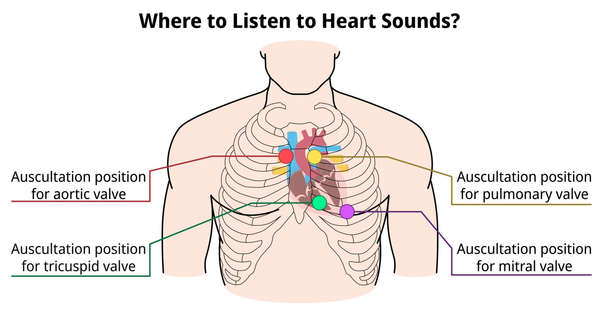
Choice A reason: The tricuspid valve is typically auscultated at the fourth or fifth intercostal space along the left lower sternal border. This anatomical site allows for the best transmission of sounds produced by the closure of the right atrioventricular valve, which separates the right atrium from the right ventricle during the cardiac cycle.
Choice B reason: The aortic valve is best assessed at the second intercostal space, just to the right of the sternal border. This location, known as the aortic area, facilitates the detection of the S2 heart sound and specific murmurs such as aortic stenosis, where blood flow is ejected into the ascending aorta.
Choice C reason: The mitral valve, or bicuspid valve, is located at the fifth intercostal space at the left midclavicular line, which corresponds to the cardiac apex. This site provides the most direct acoustic window to the left ventricle, making it the primary location for assessing the S1 heart sound and apical pulse.
Getty Images
Choice D reason: The pulmonic valve is auscultated at the second intercostal space, immediately to the left of the sternal border. This area is designated for monitoring the closure of the semilunar valve between the right ventricle and the pulmonary artery, where the pulmonic component of the second heart sound is loudest.
Correct Answer is A
Explanation
Choice A reason: A blowing or swooshing sound is the classic description of a heart murmur, which indicates turbulent blood flow. In a client with a history of rheumatic fever, this is frequently caused by mitral regurgitation (an incompetent valve) or mitral stenosis (a narrowed valve) resulting from chronic valvular scarring.
Choice B reason: While valvular dysfunction can eventually lead to heart failure and reduced cardiac output, the sound itself does not quantify the volume of blood pumped per minute. Assessing cardiac output requires more invasive monitoring or echocardiographic measurements of stroke volume and heart rate, rather than simple auscultation of a murmur.
Choice C reason: Left atrial enlargement is a common secondary consequence of mitral valve disease, especially mitral stenosis. However, the blowing sound heard during auscultation is the direct acoustic representation of the valvular defect itself (the turbulence), not the structural size of the atrium, which is better visualized through an echocardiogram.
Choice D reason: Increased pulmonary artery pressure, or pulmonary hypertension, typically manifests as a loud or accentuated S2 heart sound (specifically the P2 component). While severe mitral valve disease can lead to pulmonary hypertension over time, the "blowing, swooshing" sound at the apex specifically identifies the valvular turbulence rather than the pressure dynamics.
Whether you are a student looking to ace your exams or a practicing nurse seeking to enhance your expertise , our nursing education contents will empower you with the confidence and competence to make a difference in the lives of patients and become a respected leader in the healthcare field.
Visit Naxlex, invest in your future and unlock endless possibilities with our unparalleled nursing education contents today