The nurse is preparing to auscultate for heart sounds on a client. Which of the following technique should be used by the nurse?
Listening for all possible sounds at a time at each specified area.
Listening to the sounds at the aortic, tricuspid, pulmonic, and mitral areas.
Listening to the sounds only at the site where the apical pulse is felt to be the strongest.
Listening by inching the stethoscope in a rough Z pattern, from the base of the heart across and down, then over to the apex.
The Correct Answer is D
A. Listening for all possible sounds at a time at each specified area: This approach does not allow for specific localization of different heart sounds and murmurs, making it difficult to accurately assess the heart's condition.
B. Listening to the sounds at the aortic, tricuspid, pulmonic, and mitral areas: This option is close but lacks the systematic approach of method D. Listening at specific anatomical locations (aortic, tricuspid, pulmonic, mitral) is important, but the Z pattern allows for thorough coverage and precise localization of any abnormal sounds.
C. Listening to the sounds only at the site where the apical pulse is felt to be the strongest: This method does not cover all the important auscultation sites on the heart and may miss significant findings.
D. Listening by inching the stethoscope in a rough Z pattern, from the base of the heart across and down, then over to the apex: This technique involves a systematic approach where the nurse listens at specific locations in a structured manner, ensuring comprehensive coverage of the heart sounds and murmurs.
Nursing Test Bank
Naxlex Comprehensive Predictor Exams
Related Questions
Correct Answer is D
Explanation
A. The eyes converge to focus on the light.
This statement refers to the convergence reflex, where both eyes move medially (towards each other) to maintain single binocular vision when focusing on a near object. It is not related to the pupillary light reflex, which involves changes in pupil size in response to light.
B. The eye focuses the image in the center of the pupil.
This choice does not accurately describe the pupillary light reflex. The pupillary light reflex involves constriction of the pupil in response to light, not focusing an image in the center of the pupil.
C. Dilation of both pupils occurs in response to bright light.
This statement is incorrect. In response to bright light, the pupils should constrict, not dilate. Dilation of pupils in bright light could indicate an abnormal response, such as in cases of certain neurological conditions or drug use.
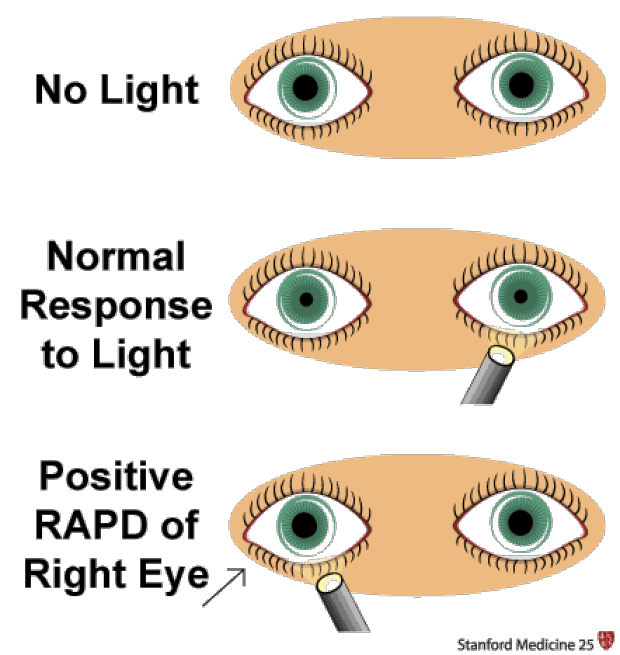
D. Constriction of both pupils occurs in response to bright light.
This choice is correct. In the pupillary light reflex, both pupils constrict when exposed to bright light. This response is a protective mechanism to prevent excessive light from entering the eyes, ensuring optimal visual acuity.
Correct Answer is ["A","B","C","D","E"]
Explanation
A. Use of accessory muscles
Explanation: Using accessory muscles during breathing indicates increased effort to breathe, which can be a sign of respiratory distress. It suggests that the client is having difficulty breathing and is using additional muscles to aid in the process. This finding should be reported to the practitioner for further evaluation.
B. Nail bed greater than 160 degrees
Explanation: A nail bed angle greater than 160 degrees, also known as clubbing, is an abnormal finding and can be associated with chronic respiratory or cardiovascular conditions. It may indicate insufficient oxygenation and should be reported to the practitioner for evaluation.
C. Circumoral cyanosis
Explanation: Circumoral cyanosis, which is a bluish discoloration around the mouth, indicates inadequate oxygenation. It can be a sign of respiratory or cardiac problems and should be reported to the practitioner for further assessment and intervention.
D. Pursed lip breathing
Explanation: Pursed lip breathing is a technique often used by individuals with respiratory difficulties to improve oxygen exchange. However, if it's observed in a person who does not normally use this technique, it could indicate respiratory distress and should be reported to the practitioner for evaluation.
E. Anteroposterior-to-transverse diameter of 1:1
Explanation: An anteroposterior-to-transverse diameter of 1:1 (also known as barrel chest) is an abnormal finding often associated with chronic obstructive pulmonary disease (COPD). It suggests overinflation of the lungs and can impair effective breathing. This finding should be reported to the practitioner for further evaluation.
Whether you are a student looking to ace your exams or a practicing nurse seeking to enhance your expertise , our nursing education contents will empower you with the confidence and competence to make a difference in the lives of patients and become a respected leader in the healthcare field.
Visit Naxlex, invest in your future and unlock endless possibilities with our unparalleled nursing education contents today