The view of this anatomical model is from what direction?
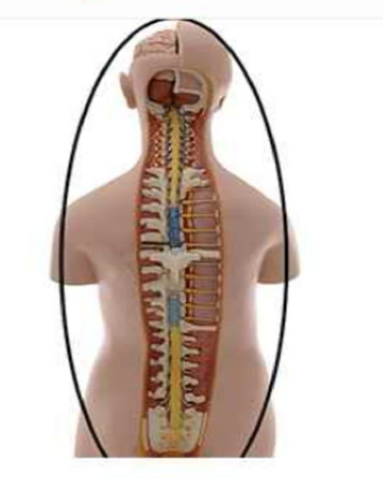
Anterior
Posterior
Lateral
Superior
The Correct Answer is B
Anatomical orientation is established by standardized planes and viewpoints, which allow clinicians and students to consistently describe the location of structures. A posterior view refers to looking at the body or an organ from the back. In the image, the model displays the vertebral column and the spinal cord exposed from the rear, which is the standard vantage point for identifying dorsal structures like the vertebral arches, spinous processes, and the posterior surface of the brainstem.
A. The anterior view refers to looking at the body from the front. If the model were viewed from the anterior, we would see the face, the chest, and the anterior surfaces of the vertebral bodies, rather than the spinal canal and the posterior vertebral elements exposed in this view.
B. The posterior view is the correct answer. This perspective shows the back of the anatomical model, revealing the spinal cord as it resides within the vertebral canal, which is accessed posteriorly by removing the laminae of the vertebrae. This orientation is essential for studying the dorsal aspects of the central nervous system.
C. A lateral view would show the model from the side. From this perspective, the structures would be seen in profile, allowing for the observation of the curvature of the spine (cervical, thoracic, and lumbar lordosis/kyphosis) and the side profiles of the vertebrae and brain, which is not the orientation presented here.
D. A superior view, or "bird's-eye view," involves looking down at the top of the model. This orientation would show the crown of the head and the cross-section of the upper neck, but it would not reveal the length of the spinal column and the central nervous system as shown in the provided image.
Nursing Test Bank
Naxlex Comprehensive Predictor Exams
Related Questions
Correct Answer is B
Explanation
Mitosis is a tightly regulated process of somatic cell division that ensures equal distribution of genetic material into two daughter cells. It is divided into distinct phases: prophase, prometaphase, metaphase, anaphase, and telophase. Each phase is characterized by specific chromosomal and spindle apparatus changes. The alignment of chromosomes at the cell’s equatorial plane is a key checkpoint ensuring proper attachment of spindle fibers before separation occurs.
A. Prophase: Prophase is the first true stage of mitosis where chromatin condenses into visible chromosomes, each consisting of two sister chromatids joined at the centromere. The nuclear envelope begins to break down, and the mitotic spindle starts forming from centrosomes. However, chromosomes are not yet aligned at the center of the cell during this phase. Prophase represents preparation for alignment rather than the alignment stage itself.
B. Metaphase: Chromosomes align along the metaphase plate, which is the equatorial plane of the cell. Spindle fibers originating from opposite poles attach to the kinetochores at the centromeres of each chromosome. This alignment ensures that each sister chromatid is properly connected to opposite spindle poles. This stage is critical for accurate chromosome segregation and serves as a major checkpoint before anaphase begins.
C. Anaphase: Anaphase occurs after metaphase and is characterized by the separation of sister chromatids. The centromeres split, and spindle fibers shorten, pulling chromatids toward opposite poles of the cell. At this stage, chromosomes are actively moving away from the center rather than aligning there. Anaphase represents the separation phase, not alignment.
D. Prometaphase: Prometaphase is the transitional phase between prophase and metaphase. During this stage, the nuclear envelope fully disintegrates, allowing spindle fibers to attach to kinetochores on chromosomes. Chromosomes begin to move toward the center of the cell but have not yet achieved stable alignment at the metaphase plate. Prometaphase precedes the actual alignment seen in metaphase.
Correct Answer is B
Explanation
Tear fluid is a protective secretion produced by the lacrimal apparatus that maintains the health of the ocular surface. It is composed of water, electrolytes, mucins, lipids, and several antimicrobial substances. Among these components, lysozyme is a key innate immune enzyme that helps defend the eye against microbial invasion. Its presence is essential because the cornea is avascular and relies heavily on tears for immune protection.
A. It buffers tear pH to maintain ocular surface stability: lysozyme does not function as a pH buffer. Tear pH is primarily maintained by bicarbonate ions and other buffering systems within the aqueous component of tears. These systems help stabilize the ocular environment for optimal enzyme activity and epithelial cell function. Lysozyme instead plays a direct antimicrobial role by targeting bacterial structures, not acid-base balance.
B. It breaks down bacterial cell walls to prevent infection: lysozyme is an antibacterial enzyme that hydrolyzes peptidoglycan, a key structural component of bacterial cell walls, especially in gram-positive organisms. By breaking the β(1→4) glycosidic bonds between N-acetylmuramic acid and N-acetylglucosamine, it weakens bacterial cell integrity, leading to osmotic lysis. This enzymatic activity provides a first-line defense mechanism on the ocular surface. It is a crucial component of innate immunity in tear secretions.
C. It increases tear viscosity to improve lubrication: tear viscosity is primarily regulated by mucins secreted by goblet cells in the conjunctiva. These glycoproteins help stabilize the tear film and enhance lubrication across the corneal surface. Lysozyme does not contribute to the physical properties of tear consistency. Its role is enzymatic defense rather than lubrication or mechanical stabilization.
D. It stimulates tear production from the lacrimal gland: tear production is controlled by neural stimulation of the lacrimal gland, primarily through parasympathetic fibers of the facial nerve (cranial nerve VII). Reflex pathways triggered by irritation or emotional stimuli activate secretion. Lysozyme is not a signaling molecule and does not regulate lacrimal gland activity. Instead, it is a component released within the tears produced by the gland.
Whether you are a student looking to ace your exams or a practicing nurse seeking to enhance your expertise , our nursing education contents will empower you with the confidence and competence to make a difference in the lives of patients and become a respected leader in the healthcare field.
Visit Naxlex, invest in your future and unlock endless possibilities with our unparalleled nursing education contents today