Identify the muscle indicated by the leader line in the image below.
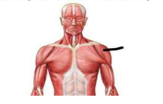
Trapezius
Biceps brachii
Deltoid
Latissimus dorsi
The Correct Answer is C
The marked structure is the deltoid muscle, a large, thick, triangular muscle covering the shoulder joint and forming the rounded contour of the shoulder. It originates from the lateral third of the clavicle, the acromion, and the spine of the scapula, and inserts on the deltoid tuberosity of the humerus. The deltoid is the primary abductor of the arm at the glenohumeral joint, especially beyond the initial 15 degrees initiated by the supraspinatus. It is also involved in flexion, extension, and rotation of the shoulder depending on the muscle fibers activated.
A. Trapezius: The trapezius is a large, superficial muscle of the upper back extending from the occipital bone to the lower thoracic vertebrae and laterally to the scapula and clavicle. It functions in scapular elevation, retraction, depression, and rotation, contributing to posture and shoulder stabilization. Unlike the deltoid, it does not act directly on the humerus or produce shoulder abduction.
B. Biceps brachii: The biceps brachii is located in the anterior compartment of the upper arm and has two heads originating from the scapula. It primarily functions in elbow flexion and forearm supination. It is not a shoulder muscle and does not form the rounded contour of the shoulder like the deltoid.
C. Deltoid: The deltoid is a multipennate muscle covering the lateral shoulder, forming its rounded contour. It abducts the arm at the shoulder joint and assists in flexion, extension, and rotation depending on fiber orientation. It originates from the clavicle, acromion, and scapular spine and inserts on the humerus. Its superficial position and shoulder-covering shape make it the correct identification.
D. Latissimus dorsi: The latissimus dorsi is a broad, flat muscle of the back that extends from the lower thoracic spine, lumbar fascia, and iliac crest to the humerus. It functions in shoulder extension, adduction, and internal rotation. Compared to the deltoid, it is located posteriorly and inferiorly and does not form the shoulder’s rounded contour.
Nursing Test Bank
Naxlex Comprehensive Predictor Exams
Related Questions
Correct Answer is C
Explanation
The skin contains specialized structures that support protection, thermoregulation, and sensation. Among these are the arrector pili muscles, which are small bundles of smooth muscle found within the dermis. These muscles play a role in thermoregulation and emotional responses by contracting to produce “goosebumps.” Their anatomical attachment is essential for their function in altering hair position on the skin surface.
A. Epidermis: The epidermis is the outermost layer of the skin composed primarily of keratinized stratified squamous epithelium. It serves as a protective barrier against environmental damage, pathogens, and water loss. It does not contain smooth muscle or serve as an attachment site for muscular structures. Arrector pili muscles are not connected to the epidermis.
B. Sebaceous glands: Sebaceous glands are exocrine glands associated with hair follicles that secrete sebum to lubricate the skin and hair. While they are located near arrector pili muscles, they are not the primary attachment site of these muscles. The arrector pili may have some structural relationship with the follicle-sebaceous unit, but their main insertion is into the hair follicle itself.
C. Hair follicles: arrector pili muscles are directly attached to hair follicles. These smooth muscles extend from the dermis and insert into the connective tissue sheath surrounding the hair follicle. When they contract, they pull the follicle into an upright position, causing hair to stand erect (piloerection). This also helps trap air for thermal insulation and is involved in the “fight-or-flight” response.
D. Nail beds: The nail bed is the skin beneath the fingernails or toenails and is involved in supporting nail growth and attachment. It consists of specialized epidermal and dermal structures that contribute to nail formation. It has no association with hair follicles or arrector pili muscles. It is anatomically unrelated to the function or attachment of these muscles.
Correct Answer is C
Explanation
The marked structure is the biceps brachii, a prominent muscle of the anterior compartment of the upper arm. It has two heads (long and short) originating from the scapula and inserting on the radial tuberosity of the radius. It crosses both the shoulder and elbow joints, making it a biarticular muscle involved in both shoulder stabilization and forearm movement. Its primary physiological functions include elbow flexion and forearm supination, especially when the forearm is in a flexed position.
A. Triceps brachii: The triceps brachii is located in the posterior compartment of the upper arm and is the primary extensor of the elbow joint. It has three heads (long, lateral, and medial) and inserts on the olecranon process of the ulna. Unlike the biceps brachii, it functions to straighten the elbow rather than flex it. Its posterior position and extensor role differentiate it from the anteriorly located biceps.
B. Brachialis: The brachialis lies deep to the biceps brachii in the anterior arm and is the strongest pure flexor of the elbow joint. It originates from the distal half of the humerus and inserts on the ulna. Unlike the biceps brachii, it does not cross the shoulder joint or contribute to forearm supination. It is a deeper muscle and not typically visible as the prominent anterior arm contour.
C. Biceps brachii: The biceps brachii is a superficial anterior arm muscle with two heads originating from the scapula (supraglenoid tubercle and coracoid process). It inserts on the radial tuberosity and via the bicipital aponeurosis into the forearm fascia. It functions in elbow flexion and powerful forearm supination. Its superficial position and characteristic bulge in the anterior upper arm make it the correct identification.
D. Brachioradialis: The brachioradialis is a forearm muscle located on the lateral aspect of the forearm. It originates from the lateral supracondylar ridge of the humerus and inserts on the distal radius. It assists in elbow flexion, especially in mid-pronation/supination positions. Unlike the biceps brachii, it is primarily a forearm muscle and does not create the prominent anterior upper arm contour.
Whether you are a student looking to ace your exams or a practicing nurse seeking to enhance your expertise , our nursing education contents will empower you with the confidence and competence to make a difference in the lives of patients and become a respected leader in the healthcare field.
Visit Naxlex, invest in your future and unlock endless possibilities with our unparalleled nursing education contents today