When auscultating Erb’s point using a systematic Z-pattern approach, where should the nurse place the stethoscope?
Right sternal border at the second intercostal space
Left sternal border at the fourth intercostal space
Left sternal border at the third intercostal space
Left midclavicular line at the fifth intercostal space
The Correct Answer is C
Choice A reason: The right sternal border at the second intercostal space is the anatomical landmark for the aortic valve auscultation site. It is distinct from Erb's point and is specifically used to listen for sounds generated by blood flow through the aortic valve into the ascending aorta.
Choice B reason: The left sternal border at the fourth intercostal space is the standard landmark for the tricuspid valve auscultation site. This position allows the nurse to best hear sounds originating from the tricuspid valve as the leaflets close during ventricular systole, rather than the primary point for Erb's.
Choice C reason: Erb's point is traditionally located at the left sternal border in the third intercostal space. It is a critical anatomical site where the sounds of both the aortic and pulmonic valves are often heard with equal intensity, making it an optimal location for detecting cardiac murmurs.
Choice D reason: The left midclavicular line at the fifth intercostal space is the location of the mitral valve, also known as the apex of the heart or the point of maximal impulse. It is where the first heart sound is typically heard best and is not the location for Erb's point.
Nursing Test Bank
Naxlex Comprehensive Predictor Exams
Related Questions
Correct Answer is B
Explanation
Choice A reason: While the cuff must be placed above the antecubital space, a fixed 5 cm distance is not the universal standard for all arm lengths. The priority is to ensure the bladder of the cuff is centered over the brachial artery and does not interfere with the placement of the stethoscope or sensor.
Choice B reason: The artery indicator (often an arrow on the cuff) must be correctly aligned with the anatomical path of the brachial artery. Misalignment can lead to inaccurate pressure readings, as the sensor or stethoscope will not be directly over the point of pulse detection, resulting in an erroneous systolic or diastolic value.
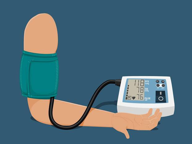
Choice C reason: Elevating the arm above the level of the heart will produce a falsely low blood pressure reading due to the effects of gravity on venous return and hydrostatic pressure. The arm must be supported and positioned at the level of the heart to ensure an accurate measurement that reflects central arterial pressure.
Choice D reason: Selecting a cuff that covers only 50% of the upper arm is incorrect; a cuff that is too small (narrow) will lead to a falsely high blood pressure reading. The standard requirement is for the cuff bladder to cover at least 80% of the arm circumference and 40% of the arm length.
Correct Answer is A
Explanation
Choice A reason: The mitral valve, also known as the bicuspid valve, is an atrioventricular valve located between the left atrium and the left ventricle. Its primary physiological function is to ensure unidirectional blood flow by closing tightly during ventricular systole, thereby preventing regurgitation of high-pressure blood back into the left atrium.
Choice B reason: The tricuspid valve is situated between the right atrium and the right ventricle. It functions to prevent the retrograde flow of deoxygenated blood into the right atrium during right ventricular contraction. It is anatomically positioned on the right side of the heart and does not manage left-sided pressures.
Choice C reason: The aortic valve is a semilunar valve located between the left ventricle and the aorta. Its function is to prevent blood from flowing back from the systemic circulation into the left ventricle during ventricular diastole. It does not regulate blood flow between the atrial and ventricular chambers.
Choice D reason: The pulmonary valve is a semilunar valve positioned between the right ventricle and the pulmonary artery. It serves to prevent the backflow of deoxygenated blood from the pulmonary vasculature into the right ventricle during the relaxation phase of the cardiac cycle, maintaining efficient forward flow to the lungs.
Whether you are a student looking to ace your exams or a practicing nurse seeking to enhance your expertise , our nursing education contents will empower you with the confidence and competence to make a difference in the lives of patients and become a respected leader in the healthcare field.
Visit Naxlex, invest in your future and unlock endless possibilities with our unparalleled nursing education contents today