Which structure is indicated by the highlight and leader line?
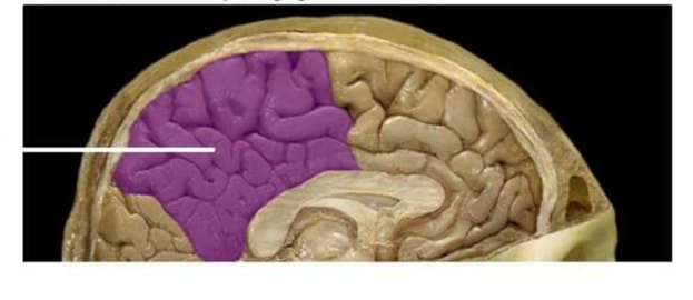
Frontal lobe
Temporal lobe
Occipital lobe
Parietal lobe
The Correct Answer is D
The marked structure is the parietal lobe, a major division of the cerebral cortex located superiorly in the brain between the frontal and occipital lobes. It lies posterior to the central sulcus and anterior to the occipital lobe, forming a significant portion of the superior and lateral aspects of each cerebral hemisphere. The parietal lobe is primarily responsible for processing somatosensory information such as touch, pressure, temperature, and pain. It also integrates sensory input to support spatial awareness, body orientation, and proprioception.
A. Frontal lobe: The frontal lobe is located anteriorly in the cerebrum, in front of the central sulcus. It is responsible for executive functions such as decision-making, judgment, personality, voluntary motor control, and speech production via Broca’s area. Compared to the parietal lobe, it is more anterior and not primarily involved in somatosensory integration or spatial processing.
B. Temporal lobe: The temporal lobe is located on the lateral aspect of the brain beneath the lateral sulcus. It is primarily involved in auditory processing, language comprehension (Wernicke’s area), and memory formation. Unlike the parietal lobe, it does not process primary somatosensory input or spatial body awareness.
C. Occipital lobe: The occipital lobe is located at the posterior pole of the brain and is the primary center for visual processing. It receives and interprets visual stimuli from the retina via the optic pathways. Compared to the parietal lobe, it is more posterior and specialized for vision rather than somatic sensation or spatial integration.
D. Parietal lobe: The parietal lobe is positioned superiorly and centrally on the cerebral hemispheres, posterior to the frontal lobe and anterior to the occipital lobe. It contains the primary somatosensory cortex located in the postcentral gyrus, which processes tactile and proprioceptive input from the body. It integrates sensory information to support spatial awareness, body positioning, and coordination of movement. Its location and function correspond to the marked region.
Nursing Test Bank
Naxlex Comprehensive Predictor Exams
Related Questions
Correct Answer is A
Explanation
The human body is organized into several major anatomical cavities that house and protect delicate internal organs. These cavities are lined by specific membranes and are categorized based on their location within the body's structural framework. The cranial cavity is a specialized dorsal space that acts as the protective bony container for the central nervous system, ensuring the brain remains shielded from external trauma.
A. The cranial cavity, also known as the endocranium, is the space enclosed by the bones of the skull. It directly houses the brain, the meninges, and the cerebrospinal fluid that cushions the brain. This cavity is continuous with the vertebral canal, forming the dorsal body cavity, which is the primary site of protection for the central nervous system.
B. The thoracic cavity is located in the upper trunk of the body, protected by the rib cage and the sternum. It houses vital organs such as the heart, lungs, esophagus, and trachea. It is separated from the abdominal cavity below by the diaphragm and is completely separate from the cranial cavity shown in the diagram.
C. The abdominal cavity occupies the upper portion of the abdominopelvic cavity. It contains the majority of the digestive organs, including the stomach, liver, intestines, and kidneys. Because it is located in the trunk and holds visceral organs associated with digestion and metabolism, it bears no anatomical relationship to the cranial cavity.
D. The pelvic cavity is the lowermost portion of the abdominopelvic cavity, located within the bony pelvis. It contains the urinary bladder, reproductive organs, and the terminal portions of the gastrointestinal tract. Like the abdominal cavity, it is positioned far from the head and is not involved in housing the brain.
Correct Answer is B
Explanation
Tear fluid is a protective secretion produced by the lacrimal apparatus that maintains the health of the ocular surface. It is composed of water, electrolytes, mucins, lipids, and several antimicrobial substances. Among these components, lysozyme is a key innate immune enzyme that helps defend the eye against microbial invasion. Its presence is essential because the cornea is avascular and relies heavily on tears for immune protection.
A. It buffers tear pH to maintain ocular surface stability: lysozyme does not function as a pH buffer. Tear pH is primarily maintained by bicarbonate ions and other buffering systems within the aqueous component of tears. These systems help stabilize the ocular environment for optimal enzyme activity and epithelial cell function. Lysozyme instead plays a direct antimicrobial role by targeting bacterial structures, not acid-base balance.
B. It breaks down bacterial cell walls to prevent infection: lysozyme is an antibacterial enzyme that hydrolyzes peptidoglycan, a key structural component of bacterial cell walls, especially in gram-positive organisms. By breaking the β(1→4) glycosidic bonds between N-acetylmuramic acid and N-acetylglucosamine, it weakens bacterial cell integrity, leading to osmotic lysis. This enzymatic activity provides a first-line defense mechanism on the ocular surface. It is a crucial component of innate immunity in tear secretions.
C. It increases tear viscosity to improve lubrication: tear viscosity is primarily regulated by mucins secreted by goblet cells in the conjunctiva. These glycoproteins help stabilize the tear film and enhance lubrication across the corneal surface. Lysozyme does not contribute to the physical properties of tear consistency. Its role is enzymatic defense rather than lubrication or mechanical stabilization.
D. It stimulates tear production from the lacrimal gland: tear production is controlled by neural stimulation of the lacrimal gland, primarily through parasympathetic fibers of the facial nerve (cranial nerve VII). Reflex pathways triggered by irritation or emotional stimuli activate secretion. Lysozyme is not a signaling molecule and does not regulate lacrimal gland activity. Instead, it is a component released within the tears produced by the gland.
Whether you are a student looking to ace your exams or a practicing nurse seeking to enhance your expertise , our nursing education contents will empower you with the confidence and competence to make a difference in the lives of patients and become a respected leader in the healthcare field.
Visit Naxlex, invest in your future and unlock endless possibilities with our unparalleled nursing education contents today