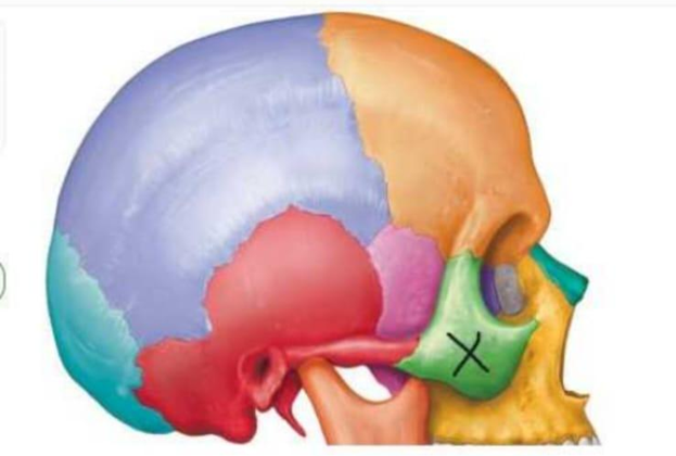
Which structure is indicated by the X in the image below?
Zygomatic bone
Maxilla
Temporal bone
Sphenoid bone
The Correct Answer is A
The human skull is a complex assembly of multiple bones joined by immovable sutures, providing protection for the brain and structural support for the facial skeleton. The lateral aspect of the skull features the zygomatic bone, often referred to as the "cheekbone," which forms a significant portion of the lateral orbital wall and the zygomatic arch. Recognizing these anatomical landmarks is essential for understanding facial trauma, surgical approaches, and the structural integrity of the cranium.
A. The zygomatic bone is the correct identification for the structure marked with an "X." This paired bone forms the prominence of the cheek, the lateral orbital margin, and part of the floor of the orbit. It articulates with the temporal, frontal, sphenoid, and maxillary bones to create the robust architecture of the midface.
B. The maxilla is the upper jaw bone that forms the central part of the face, including the nasal aperture and the majority of the hard palate. While it is located adjacent to the zygomatic bone, it is positioned more medially and inferiorly. It is not the bone marked by the "X," which is clearly located on the lateral prominence of the cheek.
C. The temporal bone is a large, complex bone located on the side and base of the skull, which houses the structures of the inner ear. It articulates with the zygomatic process to form the zygomatic arch, but it is situated posterior to the zygomatic bone. The structure marked with the "X" is the anterior cheek prominence, not the lateral cranial bone.
D. The sphenoid bone is a complex, butterfly-shaped bone situated at the base of the cranium. It acts as a keystone, articulating with almost all other cranial bones. While part of the greater wing of the sphenoid is visible on the lateral skull, it is located deeper within the skull anatomy and is not the superficial "cheekbone" structure indicated in the image.
Nursing Test Bank
Naxlex Comprehensive Predictor Exams
Related Questions
Correct Answer is B
Explanation
The marked structure is the pupil, the central circular opening within the iris through which light enters the eye. Although commonly perceived as a black structure, the pupil is actually an aperture rather than a physical tissue. Its diameter changes continuously in response to light intensity and autonomic nervous system stimulation through the actions of the sphincter pupillae and dilator pupillae muscles located within the iris. Regulation of pupil size is essential for controlling the amount of light reaching the retina and optimizing visual acuity under varying environmental conditions.
A. Cornea: The cornea is the transparent, avascular anterior portion of the fibrous tunic of the eye that covers the iris, pupil, and anterior chamber. It provides approximately two-thirds of the eye’s refractive power by bending incoming light toward the retina. Unlike the pupil, the cornea is a physical structure composed of specialized layers of tissue and does not constrict or dilate in response to light.
B. Pupil: The pupil is the circular opening located at the center of the iris and serves as the gateway through which light enters the eye. Its size changes through pupillary constriction (miosis) and dilation (mydriasis), allowing regulation of retinal light exposure. Parasympathetic stimulation causes constriction, whereas sympathetic stimulation causes dilation. Because the marked structure is the small circular opening that changes diameter in response to light, it is the pupil.
C. Lens: The lens is a transparent, biconvex structure located directly posterior to the iris and pupil. It functions by altering its shape during accommodation to focus light rays precisely onto the retina for near and distant vision. Unlike the pupil, the lens is a solid anatomical structure and does not change size to regulate light entry. Its role is optical focusing rather than light regulation.
D. Iris: The iris is the pigmented, circular structure surrounding the pupil and responsible for determining eye color. It contains smooth muscle fibers arranged as the sphincter pupillae and dilator pupillae muscles, which control pupil diameter. While the iris performs the mechanical action that changes pupil size, the opening that actually constricts and dilates is the pupil itself. Therefore, the iris surrounds the marked structure but is not the structure being identified.
Correct Answer is A
Explanation
Muscle tissue in the human body is classified into three types based on structure and control mechanisms: skeletal, smooth, and cardiac muscle. These tissues differ in their microscopic organization, location, and mode of nervous system regulation. Voluntary control refers to conscious activation via the somatic nervous system, while involuntary control is regulated by the autonomic nervous system and intrinsic pacemaker activity. Understanding these differences is essential for distinguishing body systems involved in movement versus automatic physiological functions.
A. Smooth muscle tissue and cardiac muscle tissue: both smooth and cardiac muscle tissues are under involuntary control. Smooth muscle, found in structures such as blood vessels, the gastrointestinal tract, and respiratory passages, is regulated by the autonomic nervous system and local chemical signals. Cardiac muscle, located in the heart, is also involuntary and has intrinsic rhythmic activity controlled by the sinoatrial node, with modulation from autonomic inputs. Both muscle types function automatically without conscious control.
B. Skeletal muscle tissue and smooth muscle tissue: skeletal muscle is under voluntary control, not involuntary control. Skeletal muscles are innervated by the somatic nervous system and require conscious effort for activation, such as walking or lifting objects. While smooth muscle is involuntary, pairing it with skeletal muscle makes this option incorrect overall.
C. Smooth muscle tissue, skeletal muscle tissue, and cardiac muscle tissue: This option includes all three muscle types, but skeletal muscle is voluntary. Although smooth and cardiac muscles are involuntary, skeletal muscle is consciously controlled. Therefore, not all listed muscle types are involuntary.
D. Skeletal muscle tissue only: skeletal muscle is entirely voluntary and controlled by the somatic nervous system. It is responsible for conscious movements such as locomotion and posture maintenance. It does not function automatically or independently of conscious control. Therefore, it cannot be classified as involuntary muscle tissue.
Whether you are a student looking to ace your exams or a practicing nurse seeking to enhance your expertise , our nursing education contents will empower you with the confidence and competence to make a difference in the lives of patients and become a respected leader in the healthcare field.
Visit Naxlex, invest in your future and unlock endless possibilities with our unparalleled nursing education contents today