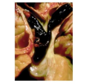
A 56-year-old man who has been at home recovering following an appendectomy, collapses after rushing out of bed to answer the telephone. Autopsy findings are shown (see image). This thromboembolus most likely originated in which of the following anatomic sites?
Left ventricle of heart
Leg vein
Pulmonary artery
Right ventricle of heart
Thoracic aorta
The Correct Answer is B
A. Left ventricle of heart: Thrombi in the left ventricle are typically associated with myocardial infarction or cardiomyopathy and can lead to systemic arterial emboli (e.g., stroke or limb ischemia). They do not travel to the pulmonary arteries, so this is unlikely.
B. Leg vein: Most pulmonary emboli originate from deep vein thromboses (DVT) in the lower extremities, particularly the femoral, popliteal, or iliac veins. Dislodged thrombi travel through the venous system to the right heart and then lodge at the pulmonary artery bifurcation, producing a saddle embolus, as seen in this patient.
C. Pulmonary artery: Thrombi do not typically form in the pulmonary arteries themselves; they usually arrive there as emboli from peripheral veins. Primary pulmonary artery thrombosis is rare.
D. Right ventricle of heart: Right ventricular thrombi are uncommon and typically occur in situ due to catheter-related injury or severe right heart dysfunction. They are not the usual source of a sudden saddle pulmonary embolus.
E. Thoracic aorta: Thrombi in the aorta are part of the systemic arterial circulation and can embolize to peripheral organs, not to the pulmonary vasculature. They cannot cause pulmonary embolism.
Nursing Test Bank
Naxlex Comprehensive Predictor Exams
Related Questions
Correct Answer is B
Explanation
A. Acute left-sided heart failure:Left-sided heart failure typically presents with pulmonary congestion, dyspnea, orthopnea, and pulmonary rales. While it can eventually lead to right-sided symptoms, this patient’s presentation is dominated by right-sided signs—jugular venous distention, peripheral edema, hepatomegaly—without primary pulmonary edema, making left-sided failure less likely.
B. Cor pulmonale:Cor pulmonale is right ventricular enlargement and dysfunction caused by chronic pulmonary hypertension, often secondary to chronic lung diseases such as COPD. Features include peripheral edema, cyanosis, elevated jugular venous pressure, loud P2 due to pulmonary hypertension, hepatomegaly, and echocardiographic evidence of right ventricular dilation and hypertrophy. The patient’s history of severe COPD and chronic hypoxia strongly supports this diagnosis.
C. Pulmonary embolism:Pulmonary embolism can cause acute right heart strain and dyspnea, but it usually presents suddenly with chest pain, hemoptysis, and often without chronic signs such as peripheral edema or hepatomegaly. Echocardiography may show right ventricular dilation acutely, but chronic hypertrophy is not typical in isolated PE.
D. Dilated cardiomyopathy:Dilated cardiomyopathy affects both ventricles with progressive systolic dysfunction, leading to biventricular heart failure. While it can cause right-sided symptoms, the patient’s chronic COPD history and predominance of right-sided findings point to cor pulmonale rather than primary dilated cardiomyopathy.
Correct Answer is D
Explanation
A. Adenocarcinoma of the lung:Adenocarcinoma is the most common type of lung cancer in nonsmokers and often arises in the peripheral lung tissue. It can develop due to genetic factors, environmental exposures, or unknown causes, making it possible even in individuals with no history of smoking or secondhandsmoke.
B. Small cell carcinoma of the lung:Small cell carcinoma is strongly associated with smoking but can rarely occur in nonsmokers. While uncommon, its occurrence is not impossible in someone who has never smoked, especially if other risk factors are present.
C. Mature carcinoid tumor:Carcinoid tumors are neuroendocrine tumors that often occur in the lungs independently of smoking. They are slow-growing and can develop in individuals with no history of tobacco exposure, making them likely in nonsmokers.
D. Squamous cell carcinoma of the lung:Squamous cell carcinoma is strongly linked to cigarette smoking and chronic exposure to tobacco smoke. It typically arises in the central bronchi and is extremely rare in individuals who have never smoked or been exposed to significant secondhand smoke, making it the least likely lung cancer in this patient.
Whether you are a student looking to ace your exams or a practicing nurse seeking to enhance your expertise , our nursing education contents will empower you with the confidence and competence to make a difference in the lives of patients and become a respected leader in the healthcare field.
Visit Naxlex, invest in your future and unlock endless possibilities with our unparalleled nursing education contents today