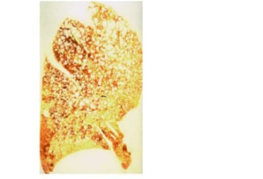
A 68-year-old man with a history of heavy smoking suffers a massive heart attack and expires. A whole-mount of the patient's lungs is examined at autopsy (shown). This patient suffered from which of the following chronic pulmonary diseases?
Atelectasis
Bronchiectasis
Chronic bronchitis
Emphysema
Pneumonia
The Correct Answer is D
A. Atelectasis: Atelectasis is collapse of lung tissue due to obstruction, compression, or surfactant deficiency. It results in decreased lung volume rather than hyperinflation and does not produce enlarged, irregular air spaces as seen in this patient.
B. Bronchiectasis: Bronchiectasis involves permanent dilation of bronchi due to chronic infection or obstruction, often associated with copious purulent sputum. It primarily affects the airways rather than alveolar structures and does not cause the centriacinar destruction or “sponge-like” hyperinflated lungs described.
C. Chronic bronchitis: Chronic bronchitis is characterized by chronic productive cough due to hyperplasia of mucus-secreting glands and airway inflammation. While it may coexist with emphysema, it does not produce enlarged alveolar air spaces or the characteristic upper-lobe centriacinar pattern.
D. Emphysema: Emphysema is a chronic obstructive pulmonary disease marked by irreversible enlargement of alveoli and destruction of alveolar walls. Centriacinar (centrilobular) emphysema predominantly is strongly associated with heavy smoking. The “sponge-like,” hyperinflated lungs with irregular air spaces are classic features.
E. Pneumonia: Pneumonia is an acute infection causing alveolar consolidation, inflammation, and exudate accumulation. It does not lead to permanent alveolar enlargement or centriacinar destruction and is inconsistent with the chronic “sponge-like” changes observed in this patient.
Nursing Test Bank
Naxlex Comprehensive Predictor Exams
Related Questions
Correct Answer is B
Explanation
A. Acute myocardial infarction:Acute myocardial infarction presents with chest pain, often radiating to the arm, neck, or jaw, and may cause hypotension if extensive. However, pain extending to the abdominal cavity and the sudden drop in blood pressure in a hypertensive patient are more characteristic of aortic dissection than myocardial infarction.
B. Dissecting aortic aneurysm:Aortic dissection typically occurs in patients with long-standing hypertension. It presents with sudden, severe chest pain radiating to the back or abdomen, sometimes described as tearing. Hypotension can develop if there is rupture into the pericardium, pleura, or retroperitoneum, making this the most likely medical emergency in this patient.
C. Pulmonary thromboembolism and infarction:Pulmonary embolism usually causes sudden dyspnea, pleuritic chest pain, tachypnea, and sometimes hemoptysis. While hypotension can occur in massive PE, the characteristic tearing chest-to-abdomen pain is not typical, making PE less likely.
D. Ruptured myocardium and hemopericardium:Rupture of the myocardium typically occurs after a large transmural infarction, leading to sudden cardiac tamponade and hypotension. Pain is usually acute and severe, but the radiating pain to the abdominal cavity is less characteristic than in aortic dissection.
E. Thrombosis of hepatic veins:Hepatic vein thrombosis (Budd-Chiari syndrome) presents with abdominal pain, hepatomegaly, and ascites. It does not cause sudden chest pain radiating to the abdomen or acute hypotension, making it unlikely in this scenario.
Correct Answer is D
Explanation
A. Atelectasis:Atelectasis is collapse of lung tissue due to obstruction, compression, or surfactant deficiency. It results in decreased lung volume rather than hyperinflation and does not produce enlarged, irregular air spaces as seen in this patient.
B. Bronchiectasis:Bronchiectasis involves permanent dilation of bronchi due to chronic infection or obstruction, often associated with copious purulent sputum. It primarily affects the airways rather than alveolar structures and does not cause the centriacinar destruction or “sponge-like” hyperinflated lungs described.
C. Chronic bronchitis:Chronic bronchitis is characterized by chronic productive cough due to hyperplasia of mucus-secreting glands and airway inflammation. While it may coexist with emphysema, it does not produce enlarged alveolar air spaces or the characteristic upper-lobe centriacinar pattern.
D. Emphysema:Emphysema is a chronic obstructive pulmonary disease marked by irreversible enlargement of alveoli and destruction of alveolar walls. Centriacinar (centrilobular) emphysema predominantly is strongly associated with heavy smoking. The “sponge-like,” hyperinflated lungs with irregular air spaces are classic features.
E. Pneumonia:Pneumonia is an acute infection causing alveolar consolidation, inflammation, and exudate accumulation. It does not lead to permanent alveolar enlargement or centriacinar destruction and is inconsistent with the chronic “sponge-like” changes observed in this patient.
Whether you are a student looking to ace your exams or a practicing nurse seeking to enhance your expertise , our nursing education contents will empower you with the confidence and competence to make a difference in the lives of patients and become a respected leader in the healthcare field.
Visit Naxlex, invest in your future and unlock endless possibilities with our unparalleled nursing education contents today