Identify the component of the heart pointed to by the line.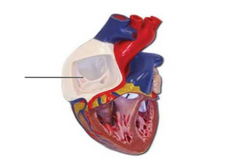
The component is
Trabecular carneae
Pulmonary semilunar valve
Papillary muscles
Pectinate muscles
Chordae tendineae
Right atrium
The Correct Answer is F
Correct answer: F.
A. Trabeculae carneae: These are irregular, muscular ridges lining the inner walls of the ventricles. They prevent suction during contraction, aid in ventricular contraction efficiency, and contribute to overall cardiac structural integrity.
B. Pulmonary semilunar valve: This valve is located between the right ventricle and pulmonary artery. It prevents backflow of blood into the ventricle during diastole and ensures unidirectional pulmonary circulation toward the lungs.
C. Papillary muscles: Papillary muscles are conical projections of ventricular myocardium that anchor chordae tendineae. During ventricular contraction, they contract to prevent inversion or prolapse of atrioventricular valves, ensuring proper unidirectional blood flow.
D. Pectinate muscles: Pectinate muscles are comb-like muscular ridges in the atrial walls, particularly prominent in the right atrium. They enhance atrial contraction, increasing blood flow into the ventricles efficiently during systole.
E. Chordae tendineae: These are thin, fibrous cords connecting atrioventricular valve leaflets to papillary muscles. They prevent valve prolapse during ventricular contraction, maintaining proper closure and unidirectional blood flow from atria to ventricles.
F. Right atrium: It is located on the superior right side of the heart and receives deoxygenated blood from the superior vena cava, inferior vena cava, and coronary sinus. It forms the right border of the heart. Physiologically, the right atrium functions as a receiving chamber that collects systemic venous blood and delivers it through the tricuspid valve into the right ventricle during atrial contraction.
G. Bicuspid valve: Also called the mitral valve, it is located between the left atrium and left ventricle. It prevents backflow into the atrium during ventricular contraction, ensuring efficient systemic circulation.
H. Fossa ovalis: This is a depression in the interatrial septum, the remnant of the fetal foramen ovale. It allowed blood to bypass the fetal lungs and normally closes after birth.
I. Left ventricle: The left ventricle pumps oxygenated blood into the aorta under high pressure. Its thick muscular wall enables strong contractions necessary to sustain systemic circulation throughout the body.
J. Interventricular septum: This thick muscular wall separates the left and right ventricles. It prevents mixing of oxygenated and deoxygenated blood and contributes to the contractile force of ventricular systole.
Nursing Test Bank
Naxlex Comprehensive Predictor Exams
Related Questions
Correct Answer is B
Explanation
Correct answer: False
The atrioventricular (AV) node, located in the lower portion of the interatrial septum near the tricuspid valve, functions as a critical electrical relay between the atria and ventricles. While the SA node sets the normal heart rhythm, the AV node limits the number of impulses transmitted to the ventricles, providing a protective delay that allows adequate ventricular filling. Under extreme SA node stimulation, impulses exceeding 220 per minute can overwhelm the AV node, resulting in ineffective ventricular contractions, reduced cardiac output, and compromised heart function. The AV node’s intrinsic conduction limits are essential for maintaining coordinated and efficient cardiac performance.
Correct Answer is F
Explanation
A. Small cardiac vein: The small cardiac vein runs along the right margin of the heart and drains blood from the right atrium and ventricle. It empties into the coronary sinus, facilitating venous return to the right atrium.
B. Coronary sinus: The coronary sinus is a large venous channel on the posterior aspect of the heart. It collects most cardiac venous blood and drains directly into the right atrium, completing coronary circulation.
C. Anterior cardiac vein: The anterior cardiac veins run along the anterior surface of the right ventricle. They bypass the coronary sinus, draining directly into the right atrium, and contribute to the venous return from the right ventricular myocardium.
D. Superior vena cava: The superior vena cava is a major systemic vein that returns deoxygenated blood from the upper body, including the head, neck, and upper limbs, directly into the right atrium of the heart.
E. Middle cardiac vein; The middle cardiac vein runs in the posterior interventricular sulcus, draining the posterior portion of both ventricles. It empties into the coronary sinus, ensuring efficient venous return from the heart’s posterior myocardium.
F. Great cardiac vein: The blood vessel highlighted in the image is the great cardiac vein located on the anterior surface of the heart. The great cardiac vein begins at the apex of the heart and eventually curves around the left side of the heart (within the coronary sulcus) to empty into the coronary sinus on the posterior side. It is the principal vein of the anterior heart.
Whether you are a student looking to ace your exams or a practicing nurse seeking to enhance your expertise , our nursing education contents will empower you with the confidence and competence to make a difference in the lives of patients and become a respected leader in the healthcare field.
Visit Naxlex, invest in your future and unlock endless possibilities with our unparalleled nursing education contents today