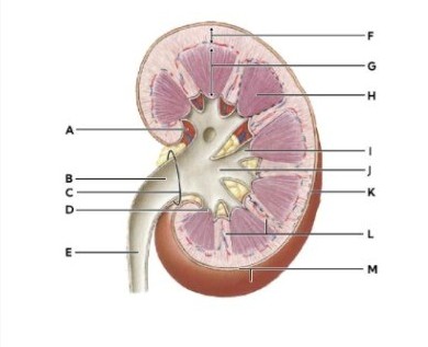
Identify the following structures by letter and make sure to number your answer for grading.
|
|
|
|
Minor calyx |
dropdown
|
|
Renal medulla |
dropdown
|
|
Renal cortex |
dropdown
|
The Correct Answer is {"dropdown-group-1":"D","dropdown-group-2":"E","dropdown-group-3":"E"}
Minor calyx (I): The minor calyx is a small, cup-shaped structure that collects urine from the renal papilla at the tip of each pyramid. It is part of the drainage system, channeling urine into the major calyces and then into the renal pelvis. Look for the small funnel-like extensions directly attached to the renal papillae. These are distinctly smaller than the major calyces.
Renal medulla(H): The renal medulla is the inner region of the kidney, composed of renal pyramids. It plays a crucial role in concentrating urine through the countercurrent mechanism in the loops of Henle and collecting ducts. Appears as darker, triangular or striated regions deeper inside the kidney, between the cortex and the renal pelvis.
Renal cortex (F): The renal cortex is the outer functional tissue of the kidney, lying just beneath the fibrous capsule. It contains glomeruli and convoluted tubules, making it the primary site of blood filtration. Appears as the lighter-colored outer rim of the kidney, surrounding the medulla.
Nursing Test Bank
Naxlex Comprehensive Predictor Exams
Related Questions
Correct Answer is B
Explanation
A. Afferent arteriole: This vessel serves as the high-resistance inflow pathway that delivers blood into the glomerular capillary tuft for filtration. It precedes the glomerulus in the renal microcirculatory sequence rather than following it. Its primary role is regulating the hydrostatic pressure within the glomerular capillaries.
B. Efferent arteriole: Blood exits the glomerular capillaries through this vessel, which maintains the high-pressure system necessary for efficient ultrafiltration. This unique portal arrangement allows for the subsequent delivery of blood to either peritubular capillaries or vasa recta. It is the immediate downstream vessel from the glomerulus.
C. Interlobar vein: These vessels are located much further downstream in the centripetal drainage system, situated between the renal pyramids. Blood must first pass through the efferent arterioles and peritubular networks before reaching the venous return. It represents a macroscopic stage of drainage rather than a direct glomerular exit.
D. Arcuate vein: These veins run along the boundary between the renal cortex and medulla, collecting blood from the cortical radiate veins. They represent a late stage in the venous exit pathway and are not in direct contact with the glomerular capillaries. Their function is to transport deoxygenated blood toward the interlobar veins.
E. Peritubular capillaries: While these vessels do receive blood from the efferent arterioles in cortical nephrons, they are not the immediate exit point from the glomerulus. The efferent arteriole serves as a necessary resistance vessel between the glomerular tuft and these low-pressure capillaries. The glomerular blood must traverse the efferent arteriole first.
Correct Answer is B
Explanation
B. False: The Proximal Convoluted Tubule (PCT) is named for its highly coiled and tortuous path through the renal cortex. This morphology increases the surface area and residence time for the reabsorption of water, ions, and nutrients. A straight path would be described as "rectus," not convoluted.
Whether you are a student looking to ace your exams or a practicing nurse seeking to enhance your expertise , our nursing education contents will empower you with the confidence and competence to make a difference in the lives of patients and become a respected leader in the healthcare field.
Visit Naxlex, invest in your future and unlock endless possibilities with our unparalleled nursing education contents today