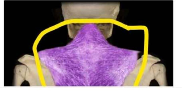
Identify the muscle that is highlighted and circled in the image below.
Trapezius
Latissimus dorsi
Deltoid
Sternocleidomastoid
The Correct Answer is A
The marked structure is the trapezius muscle, a large, superficial, triangular muscle that extends across the posterior neck and upper back. It originates from the occipital bone, ligamentum nuchae, and spinous processes of C7–T12, and inserts onto the clavicle, acromion, and spine of the scapula. The trapezius plays a major role in scapular positioning and movement, including elevation, retraction, depression, and upward rotation. It also contributes to neck extension and stabilization of the shoulder girdle.
A. Trapezius: The trapezius is a broad, superficial muscle covering the posterior neck and upper thorax, forming a diamond-shaped structure across the upper back. It controls scapular movements such as elevation (shrugging), retraction (pulling shoulders back), and rotation necessary for overhead arm activity. It is also involved in stabilizing the scapula during upper limb movement. Its extensive posterior location and attachment to the scapula and clavicle make it the correct answer.
B. Latissimus dorsi: The latissimus dorsi is a large, flat muscle of the lower back that extends to the humerus. It functions primarily in shoulder extension, adduction, and internal rotation, especially during pulling movements. Unlike the trapezius, it is located in the lower posterior trunk and does not extend into the neck region. It also does not elevate or stabilize the scapula in the same way.
C. Deltoid: The deltoid is a thick, triangular muscle covering the lateral aspect of the shoulder joint. It is responsible for abduction of the arm and contributes to flexion and extension depending on fiber segment. It is not located on the posterior back or neck, and it does not control scapular movement, unlike the trapezius.
D. Sternocleidomastoid: The sternocleidomastoid is a paired muscle located in the anterior and lateral neck. It originates from the sternum and clavicle and inserts on the mastoid process of the temporal bone. It functions in neck flexion, rotation, and lateral bending. Compared to the trapezius, it is anteriorly positioned and does not act on the scapula or upper back region.
Nursing Test Bank
Naxlex Comprehensive Predictor Exams
Related Questions
Correct Answer is C
Explanation
Neuronal communication occurs at specialized junctions called synapses, where signals are transmitted from one neuron to another. Most synapses in the nervous system are chemical synapses, relying on neurotransmitters rather than direct electrical continuity. When an action potential reaches the end of a presynaptic neuron, it triggers a cascade of events that leads to neurotransmitter release. These chemical messengers then cross the synaptic cleft and bind to receptors on the postsynaptic membrane, initiating a new electrical signal.
A. A neurotransmitter traveling from postsynaptic axons crosses the synapse to presynaptic dendrites or a cell body: neurotransmitters are released from the presynaptic neuron, not the postsynaptic neuron. Additionally, signaling does not occur in reverse direction in a typical chemical synapse. The postsynaptic neuron receives, rather than sends, the chemical signal.
B. An impulse stimulating a postsynaptic axon causes the release of neurotransmitters into a synaptic cleft: neurotransmitter release is triggered by an action potential arriving at the presynaptic axon terminal, not the postsynaptic axon. The postsynaptic neuron receives chemical signals rather than initiating them at the synapse.
C. An impulse stimulates a presynaptic axon, causing release of neurotransmitters into the synaptic cleft: when an action potential reaches the axon terminal of the presynaptic neuron, voltage-gated calcium channels open. Calcium influx triggers synaptic vesicles to fuse with the presynaptic membrane, releasing neurotransmitters into the synaptic cleft. These neurotransmitters then bind to receptors on the postsynaptic membrane, generating a new electrical response. This sequence ensures one-directional and regulated neural communication.
D. Electrical current flows directly from dendrite to dendrite across the synaptic cleft: most synapses are chemical, not electrical, and do not involve direct cytoplasmic continuity between neurons. Electrical synapses do exist (via gap junctions), but they are not the primary mechanism described here. Furthermore, synaptic transmission does not occur directly between dendrites across a cleft in typical neural signaling.
Correct Answer is C
Explanation
The skin contains specialized structures that support protection, thermoregulation, and sensation. Among these are the arrector pili muscles, which are small bundles of smooth muscle found within the dermis. These muscles play a role in thermoregulation and emotional responses by contracting to produce “goosebumps.” Their anatomical attachment is essential for their function in altering hair position on the skin surface.
A. Epidermis: The epidermis is the outermost layer of the skin composed primarily of keratinized stratified squamous epithelium. It serves as a protective barrier against environmental damage, pathogens, and water loss. It does not contain smooth muscle or serve as an attachment site for muscular structures. Arrector pili muscles are not connected to the epidermis.
B. Sebaceous glands: Sebaceous glands are exocrine glands associated with hair follicles that secrete sebum to lubricate the skin and hair. While they are located near arrector pili muscles, they are not the primary attachment site of these muscles. The arrector pili may have some structural relationship with the follicle-sebaceous unit, but their main insertion is into the hair follicle itself.
C. Hair follicles: arrector pili muscles are directly attached to hair follicles. These smooth muscles extend from the dermis and insert into the connective tissue sheath surrounding the hair follicle. When they contract, they pull the follicle into an upright position, causing hair to stand erect (piloerection). This also helps trap air for thermal insulation and is involved in the “fight-or-flight” response.
D. Nail beds: The nail bed is the skin beneath the fingernails or toenails and is involved in supporting nail growth and attachment. It consists of specialized epidermal and dermal structures that contribute to nail formation. It has no association with hair follicles or arrector pili muscles. It is anatomically unrelated to the function or attachment of these muscles.
Whether you are a student looking to ace your exams or a practicing nurse seeking to enhance your expertise , our nursing education contents will empower you with the confidence and competence to make a difference in the lives of patients and become a respected leader in the healthcare field.
Visit Naxlex, invest in your future and unlock endless possibilities with our unparalleled nursing education contents today