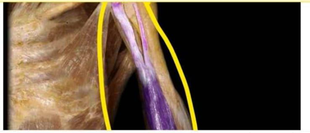
Identify the muscle that is highlighted and circled in the image below
Triceps brachii
Brachialis
Biceps brachii
Brachioradialis
The Correct Answer is C
The marked structure is the biceps brachii, a prominent muscle of the anterior compartment of the upper arm. It has two heads (long and short) originating from the scapula and inserting on the radial tuberosity of the radius. It crosses both the shoulder and elbow joints, making it a biarticular muscle involved in both shoulder stabilization and forearm movement. Its primary physiological functions include elbow flexion and forearm supination, especially when the forearm is in a flexed position.
A. Triceps brachii: The triceps brachii is located in the posterior compartment of the upper arm and is the primary extensor of the elbow joint. It has three heads (long, lateral, and medial) and inserts on the olecranon process of the ulna. Unlike the biceps brachii, it functions to straighten the elbow rather than flex it. Its posterior position and extensor role differentiate it from the anteriorly located biceps.
B. Brachialis: The brachialis lies deep to the biceps brachii in the anterior arm and is the strongest pure flexor of the elbow joint. It originates from the distal half of the humerus and inserts on the ulna. Unlike the biceps brachii, it does not cross the shoulder joint or contribute to forearm supination. It is a deeper muscle and not typically visible as the prominent anterior arm contour.
C. Biceps brachii: The biceps brachii is a superficial anterior arm muscle with two heads originating from the scapula (supraglenoid tubercle and coracoid process). It inserts on the radial tuberosity and via the bicipital aponeurosis into the forearm fascia. It functions in elbow flexion and powerful forearm supination. Its superficial position and characteristic bulge in the anterior upper arm make it the correct identification.
D. Brachioradialis: The brachioradialis is a forearm muscle located on the lateral aspect of the forearm. It originates from the lateral supracondylar ridge of the humerus and inserts on the distal radius. It assists in elbow flexion, especially in mid-pronation/supination positions. Unlike the biceps brachii, it is primarily a forearm muscle and does not create the prominent anterior upper arm contour.
Nursing Test Bank
Naxlex Comprehensive Predictor Exams
Related Questions
Correct Answer is D
Explanation
The marked structure is the lens, a transparent, biconvex, avascular structure located posterior to the iris and anterior to the vitreous body. It is suspended by zonular fibers (suspensory ligaments) attached to the ciliary body. The lens plays a critical role in vision by providing fine focusing of light onto the retina through the process of accommodation. Its curvature changes depending on whether the eye is focusing on near or distant objects, allowing precise image formation.
A. Cornea: The cornea is the transparent, dome-shaped anterior surface of the eye that provides most of the eye’s refractive power. It is the first structure that light passes through and contributes significantly to bending light toward the retina. Unlike the lens, it is external, fixed in shape, and continuous with the sclera, serving both protective and optical roles.
B. Retina: The retina is the innermost neural layer lining the posterior aspect of the eye. It contains photoreceptor cells (rods and cones) that convert light into electrical signals sent via the optic nerve to the brain. Unlike the lens, it does not focus light but instead processes visual information after image formation occurs.
C. Iris: The iris is the pigmented muscular structure located anterior to the lens and posterior to the cornea. It regulates pupil size to control the amount of light entering the eye through contraction and relaxation of its smooth muscles. Unlike the lens, it does not contribute to focusing light but only regulates light entry.
D. Lens: The lens is a transparent, flexible, biconvex structure located directly behind the iris. It fine-tunes the focusing of light rays onto the retina through accommodation, changing its curvature via the ciliary muscles. It plays a key role in sharp image formation at varying distances. Its central posterior position relative to the iris makes it the correct structure.
Correct Answer is B
Explanation
Skeletal muscle is organized into a hierarchical structure that allows coordinated contraction and force transmission. Each muscle is composed of individual muscle fibers grouped into fascicles, which are further bundled to form the entire muscle. Connective tissue layers surround each structural level to provide support, protection, and pathways for blood vessels and nerves. These layers are arranged from the deepest level surrounding individual fibers to the most superficial layer covering the entire muscle and separating it from surrounding structures.
A. Fascia → epimysium → perimysium → endomysium: This reverses the correct anatomical order from superficial to deep rather than deep to superficial. Fascia is the most superficial connective tissue layer, but epimysium lies directly over the muscle, not beneath fascia. Perimysium surrounds fascicles, and endomysium surrounds individual muscle fibers at the deepest level. Therefore, this sequence incorrectly inverts the hierarchical organization of muscle connective tissue layers.
B. Endomysium → perimysium → epimysium → fascia: This reflects the anatomical organization from deepest to most superficial layer. The endomysium surrounds individual muscle fibers, providing support and a pathway for capillaries and nerve fibers. The perimysium encloses bundles of muscle fibers called fascicles, while the epimysium surrounds the entire muscle. Finally, fascia lies most superficially, separating and encasing muscles within compartments.
C. Endomysium → epimysium → perimysium → fascia: This disrupts the correct sequence of connective tissue organization. While endomysium is correctly placed as the deepest layer, epimysium and perimysium are reversed. The perimysium must surround fascicles before the epimysium encloses the entire muscle.
D. Fascia → endomysium → perimysium → epimysium: This option begins with fascia, which is the most superficial layer rather than the deepest. It then incorrectly places endomysium beneath fascia without proper intermediate organization. Additionally, it reverses the correct order of perimysium and epimysium. This sequence does not reflect the anatomical layering of skeletal muscle connective tissues.
Whether you are a student looking to ace your exams or a practicing nurse seeking to enhance your expertise , our nursing education contents will empower you with the confidence and competence to make a difference in the lives of patients and become a respected leader in the healthcare field.
Visit Naxlex, invest in your future and unlock endless possibilities with our unparalleled nursing education contents today