Identify the highlighted structure in the image below
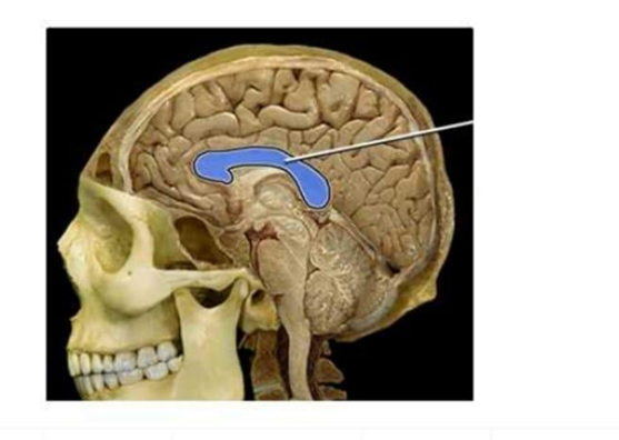
Corpus callosum
Thalamus
Hypothalamus
Cerebellum
The Correct Answer is A
The marked structure is the corpus callosum, the largest commissural fiber bundle in the brain that connects the left and right cerebral hemispheres. It is a C-shaped structure located deep within the longitudinal fissure and forms the roof of the lateral ventricles. The corpus callosum is composed of white matter tracts that enable interhemispheric communication, allowing integration of sensory, motor, and cognitive information between both hemispheres. This coordination is essential for unified brain function such as language processing, coordinated movement, and higher cognitive tasks.
A. Corpus callosum: The corpus callosum is a dense band of myelinated white matter fibers that links homologous cortical areas of both cerebral hemispheres. It facilitates rapid communication between hemispheres, ensuring coordinated motor activity, sensory integration, and cognitive processing. Anatomically, it is divided into the rostrum, genu, body, and splenium. Its midline, arched position above the lateral ventricles makes it the correct structure.
B. Thalamus: The thalamus is a paired gray matter structure located in the diencephalon superior to the brainstem and lateral to the third ventricle. It functions as the primary relay station for sensory and motor signals to the cerebral cortex. Unlike the corpus callosum, it is not a commissural fiber tract but a nuclear relay center. It is deeper and more inferior in location compared to the corpus callosum.
C. Hypothalamus: The hypothalamus is a small but critical region located inferior to the thalamus and forming the floor of the third ventricle. It regulates autonomic functions such as temperature control, hunger, thirst, endocrine regulation via the pituitary gland, and circadian rhythms. It is much smaller and more ventral than the corpus callosum and does not connect the cerebral hemispheres.
D. Cerebellum: The cerebellum is located in the posterior cranial fossa beneath the occipital lobes of the cerebrum. It is responsible for coordination of voluntary movement, balance, posture, and motor learning. Structurally, it consists of two hemispheres and a midline vermis. Unlike the corpus callosum, it is not part of the cerebrum or a commissural structure and is located posteriorly and inferiorly in the brain.
Nursing Test Bank
Naxlex Comprehensive Predictor Exams
Related Questions
Correct Answer is C
Explanation
The marked structure is the cerebellum, a major part of the hindbrain located posterior to the brainstem and inferior to the occipital lobes of the cerebrum. It consists of two hemispheres connected by the vermis and has a highly folded surface (folia) that increases its cortical area. The cerebellum is primarily responsible for coordination of voluntary movements, maintenance of posture, balance, and fine motor control. It does not initiate movement but ensures that movements are smooth, precise, and well-timed.
A. Cerebrum: The cerebrum is the largest part of the brain and consists of the cerebral hemispheres, including the frontal, parietal, temporal, and occipital lobes. It is responsible for higher cognitive functions such as reasoning, memory, language, and voluntary motor activity. Unlike the cerebellum, it is located superiorly and anteriorly in the cranial cavity and is not involved in fine motor coordination and balance regulation.
B. Brainstem: The brainstem connects the cerebrum and cerebellum to the spinal cord and consists of the midbrain, pons, and medulla oblongata. It regulates vital autonomic functions such as respiration, heart rate, and blood pressure. While it lies close to the cerebellum, it is a vertical structure inferior to the cerebrum rather than a posterior, bilobed structure like the cerebellum.
C. Cerebellum: The cerebellum is located in the posterior cranial fossa, inferior to the occipital lobes and posterior to the brainstem. It is responsible for coordinating voluntary motor activity, maintaining balance, posture, and muscle tone. It receives input from the cerebral cortex and sensory systems to fine-tune motor output. Its highly folded folia and bilateral hemispheres are characteristic features.
D. Medulla oblongata: The medulla oblongata is the lowest part of the brainstem, continuous with the spinal cord. It controls essential autonomic functions such as breathing, heart rate, and blood pressure regulation. Unlike the cerebellum, it is a narrow, tubular structure and does not have a highly folded cortical surface or function in motor coordination and balance.
Correct Answer is C
Explanation
Joints are classified based on their structure and the degree of movement they allow. Synovial joints are the most mobile type of joint in the body and are characterized by a synovial cavity, articular cartilage, and a joint capsule filled with synovial fluid. Examples include hinge, ball-and-socket, and pivot joints. In contrast, some joints are cartilaginous, where bones are united by cartilage and movement is limited. Understanding these classifications is essential for distinguishing joint anatomy and function.
A. Hinge: hinge joints are synovial joints. They allow movement in one plane, typically flexion and extension, like the elbow or interphalangeal joints. They are characterized by a synovial cavity and articular cartilage that reduce friction during movement. Therefore, hinge joints are a type of synovial joint.
B. Ball-and-socket: ball-and-socket joints are synovial joints that allow multiaxial movement, including flexion, extension, abduction, adduction, and rotation. Examples include the shoulder and hip joints. They are highly mobile due to the spherical head of one bone fitting into a cup-shaped socket. Therefore, they are classified as synovial joints.
C. Symphysis: symphysis joints are cartilaginous joints, not synovial joints. In a symphysis, bones are joined by fibrocartilage, which allows limited movement and provides strength and shock absorption. Examples include the pubic symphysis and intervertebral discs. Since they lack a synovial cavity and synovial fluid, they are not synovial joints.
D. Pivot: pivot joints are synovial joints that allow rotational movement around a single axis. A classic example is the atlantoaxial joint between the first and second cervical vertebrae, which enables head rotation. These joints have a synovial cavity and are freely movable within their functional range.
Whether you are a student looking to ace your exams or a practicing nurse seeking to enhance your expertise , our nursing education contents will empower you with the confidence and competence to make a difference in the lives of patients and become a respected leader in the healthcare field.
Visit Naxlex, invest in your future and unlock endless possibilities with our unparalleled nursing education contents today