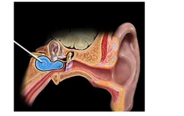
Identify the part of the ear indicated by the arrow in the image below.
Semicircular canals
Cochlea
Tympanic membrane
Auditory tube
The Correct Answer is B
The marked structure is the cochlea, a spiral-shaped, snail-like component of the inner ear located within the petrous part of the temporal bone. It is a critical organ of hearing that converts mechanical vibrations from the middle ear into electrical nerve impulses. The cochlea contains the organ of Corti, which houses hair cells that transduce sound energy into neural signals transmitted via the cochlear branch of the vestibulocochlear nerve (CN VIII). Its coiled structure allows efficient processing of different sound frequencies along its length (tonotopic organization).
A. Semicircular canals: The semicircular canals are three looped structures of the inner ear oriented in different planes (anterior, posterior, and lateral). They are part of the vestibular system and are responsible for detecting rotational movements of the head. They contain endolymph and hair cells within the ampullae that respond to angular acceleration. Unlike the cochlea, they are not involved in hearing but in balance and spatial orientation.
B. Cochlea: The cochlea is a spiral, conical structure of the inner ear responsible for auditory transduction. It contains fluid-filled chambers (scala vestibuli, media, and tympani) and the organ of Corti, which houses mechanoreceptive hair cells. Sound vibrations transmitted through the ossicles create fluid waves that stimulate these hair cells, converting mechanical energy into electrical impulses. Its distinctive coiled shape and role in hearing make it the correct identification.
C. Tympanic membrane: The tympanic membrane (eardrum) is a thin membrane separating the external ear from the middle ear. It vibrates in response to sound waves and transmits these vibrations to the ossicles (malleus, incus, stapes). Unlike the cochlea, it is not part of the inner ear and does not perform sensory transduction. It functions purely as a mechanical sound converter.
D. Auditory tube: The auditory (Eustachian) tube connects the middle ear to the nasopharynx. It equalizes air pressure across the tympanic membrane and allows drainage of middle ear secretions. It does not participate in hearing or sound transduction and is anatomically separate from the cochlea, which is located in the inner ear.
Nursing Test Bank
Naxlex Comprehensive Predictor Exams
Related Questions
Correct Answer is A
Explanation
The marked structure is the trapezius muscle, a large, superficial, triangular muscle that extends across the posterior neck and upper back. It originates from the occipital bone, ligamentum nuchae, and spinous processes of C7–T12, and inserts onto the clavicle, acromion, and spine of the scapula. The trapezius plays a major role in scapular positioning and movement, including elevation, retraction, depression, and upward rotation. It also contributes to neck extension and stabilization of the shoulder girdle.
A. Trapezius: The trapezius is a broad, superficial muscle covering the posterior neck and upper thorax, forming a diamond-shaped structure across the upper back. It controls scapular movements such as elevation (shrugging), retraction (pulling shoulders back), and rotation necessary for overhead arm activity. It is also involved in stabilizing the scapula during upper limb movement. Its extensive posterior location and attachment to the scapula and clavicle make it the correct answer.
B. Latissimus dorsi: The latissimus dorsi is a large, flat muscle of the lower back that extends to the humerus. It functions primarily in shoulder extension, adduction, and internal rotation, especially during pulling movements. Unlike the trapezius, it is located in the lower posterior trunk and does not extend into the neck region. It also does not elevate or stabilize the scapula in the same way.
C. Deltoid: The deltoid is a thick, triangular muscle covering the lateral aspect of the shoulder joint. It is responsible for abduction of the arm and contributes to flexion and extension depending on fiber segment. It is not located on the posterior back or neck, and it does not control scapular movement, unlike the trapezius.
D. Sternocleidomastoid: The sternocleidomastoid is a paired muscle located in the anterior and lateral neck. It originates from the sternum and clavicle and inserts on the mastoid process of the temporal bone. It functions in neck flexion, rotation, and lateral bending. Compared to the trapezius, it is anteriorly positioned and does not act on the scapula or upper back region.
Correct Answer is A
Explanation
The marked structure is the sacrum, a large triangular bone formed by the fusion of five sacral vertebrae located at the base of the vertebral column. It forms the posterior wall of the pelvis and articulates with the ilium at the sacroiliac joints, contributing to pelvic stability and weight transmission from the axial skeleton to the lower limbs. The sacrum also contains sacral foramina that allow passage of spinal nerves.
A. Sacrum: The sacrum is a fused bone consisting of five vertebrae (S1–S5) located between the lumbar spine and coccyx. It forms the posterior portion of the pelvic girdle and articulates with the ilium to distribute body weight during standing and movement. It contains sacral foramina for nerve passage and supports pelvic organs.
B. Coccyx: The coccyx is the small terminal segment of the vertebral column, commonly called the tailbone. It is formed by fusion of 3–5 small vertebrae and lies inferior to the sacrum. It serves as an attachment site for ligaments and pelvic floor muscles. Compared to the sacrum, it is much smaller and more distal, making it unlikely to be the marked structure.
C. Ilium: The ilium is the largest and superior portion of the hip bone, forming the broad flared structure of the pelvis. It articulates with the sacrum at the sacroiliac joint and contributes to the acetabulum. Its main role is weight-bearing and muscle attachment for the abdominal and gluteal muscles. Unlike the midline sacrum, the ilium is lateral and paired on both sides.
D. Lumbar vertebrae: The lumbar vertebrae are five large vertebrae located in the lower back above the sacrum. They provide major support for body weight and allow flexion and extension of the trunk. While they articulate directly with the sacrum, they are separate segmented bones rather than a fused triangular structure.
Whether you are a student looking to ace your exams or a practicing nurse seeking to enhance your expertise , our nursing education contents will empower you with the confidence and competence to make a difference in the lives of patients and become a respected leader in the healthcare field.
Visit Naxlex, invest in your future and unlock endless possibilities with our unparalleled nursing education contents today