Identify the highlighted muscle in the image below?
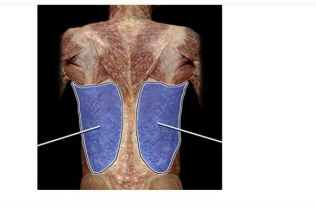
Teres major
Trapezius
Latissimus dorsi
Rhomboid major
The Correct Answer is C
The marked structure is the latissimus dorsi, a large, flat, triangular muscle that covers much of the lower posterior thorax and extends to the lateral aspect of the back. It originates from the spinous processes of T7–L5 vertebrae, thoracolumbar fascia, iliac crest, and lower ribs, and inserts into the intertubercular groove of the humerus. Its primary physiological functions include extension, adduction, and internal rotation of the humerus, making it a key muscle for pulling movements such as rowing, climbing, and swimming.
A. Teres major: The teres major is a smaller, thick muscle located inferior to the teres minor on the posterior scapula. It assists the latissimus dorsi in shoulder extension, adduction, and internal rotation. However, it is much smaller and more localized, forming part of the posterior axillary fold rather than the broad back musculature seen with the latissimus dorsi.
B. Trapezius: The trapezius is a large superficial muscle of the upper back and posterior neck that extends from the occipital region to the lower thoracic vertebrae. It controls scapular elevation, retraction, depression, and upward rotation. Unlike the latissimus dorsi, it does not act directly on the humerus and is positioned more superiorly across the neck and upper shoulders.
C. Latissimus dorsi: The latissimus dorsi is a broad, superficial muscle covering the lower and mid-back. It originates from the lower thoracic and lumbar spine, iliac crest, and ribs, and inserts into the humerus. It is responsible for powerful shoulder extension, adduction, and internal rotation, especially during activities like climbing or pulling. Its wide surface area and inferolateral back position make it the correct identification.
D. Rhomboid major: The rhomboid major is a deep muscle located between the spine and the medial border of the scapula. It functions primarily in scapular retraction and stabilization by pulling the scapula toward the vertebral column. Compared to the latissimus dorsi, it is much smaller, deeper, and does not act on the humerus.
Nursing Test Bank
Naxlex Comprehensive Predictor Exams
Related Questions
Correct Answer is D
Explanation
The wall of the eyeball is organized into three concentric layers: the outer fibrous layer, the middle vascular (uveal) layer, and the inner neural layer. The middle layer, also called the uvea, is responsible for blood supply, nourishment, and regulation of light entering the eye. It includes structures that control pupil size, lens shape, and retinal perfusion. Understanding these layers is essential for identifying ocular anatomy and related pathologies.
A. Iris: The iris is a pigmented muscular structure located in the anterior portion of the uveal tract. It contains circular (sphincter pupillae) and radial (dilator pupillae) smooth muscle fibers that regulate pupil size. This adjustment controls the amount of light entering the eye based on environmental brightness. Because it is part of the vascular middle layer, the iris is correctly included in the uvea.
B. Choroid: The choroid is a highly vascularized connective tissue layer situated between the sclera and retina. It provides oxygen and nutrient supply to the outer layers of the retina, especially the photoreceptors, which are highly metabolically active. It also absorbs excess light to prevent internal reflection within the eye. As a major component of the uveal tract, it is part of the middle eye layer.
C. Ciliary body: The ciliary body is an anterior extension of the choroid that includes the ciliary muscle and ciliary processes. It is responsible for aqueous humor production and lens accommodation by altering zonular fiber tension. This allows the lens to change shape for near and far vision focusing. Because of its vascular nature and functional integration with the iris and choroid, it is part of the middle (uveal) layer.
D. Retina: The retina is the innermost neural layer of the eye and is derived from neuroectoderm. It contains photoreceptor cells (rods and cones) that convert light energy into electrical signals through phototransduction. These signals are transmitted via bipolar and ganglion cells to the optic nerve for visual processing in the brain. Since it belongs to the inner sensory layer rather than the vascular uveal layer, it is not part of the middle eye layer.
Correct Answer is C
Explanation
The marked structure is the deltoid muscle, a large, thick, triangular muscle covering the shoulder joint and forming the rounded contour of the shoulder. It originates from the lateral third of the clavicle, the acromion, and the spine of the scapula, and inserts on the deltoid tuberosity of the humerus. The deltoid is the primary abductor of the arm at the glenohumeral joint, especially beyond the initial 15 degrees initiated by the supraspinatus. It is also involved in flexion, extension, and rotation of the shoulder depending on the muscle fibers activated.
A. Trapezius: The trapezius is a large, superficial muscle of the upper back extending from the occipital bone to the lower thoracic vertebrae and laterally to the scapula and clavicle. It functions in scapular elevation, retraction, depression, and rotation, contributing to posture and shoulder stabilization. Unlike the deltoid, it does not act directly on the humerus or produce shoulder abduction.
B. Biceps brachii: The biceps brachii is located in the anterior compartment of the upper arm and has two heads originating from the scapula. It primarily functions in elbow flexion and forearm supination. It is not a shoulder muscle and does not form the rounded contour of the shoulder like the deltoid.
C. Deltoid: The deltoid is a multipennate muscle covering the lateral shoulder, forming its rounded contour. It abducts the arm at the shoulder joint and assists in flexion, extension, and rotation depending on fiber orientation. It originates from the clavicle, acromion, and scapular spine and inserts on the humerus. Its superficial position and shoulder-covering shape make it the correct identification.
D. Latissimus dorsi: The latissimus dorsi is a broad, flat muscle of the back that extends from the lower thoracic spine, lumbar fascia, and iliac crest to the humerus. It functions in shoulder extension, adduction, and internal rotation. Compared to the deltoid, it is located posteriorly and inferiorly and does not form the shoulder’s rounded contour.
Whether you are a student looking to ace your exams or a practicing nurse seeking to enhance your expertise , our nursing education contents will empower you with the confidence and competence to make a difference in the lives of patients and become a respected leader in the healthcare field.
Visit Naxlex, invest in your future and unlock endless possibilities with our unparalleled nursing education contents today