The arrow illustrates
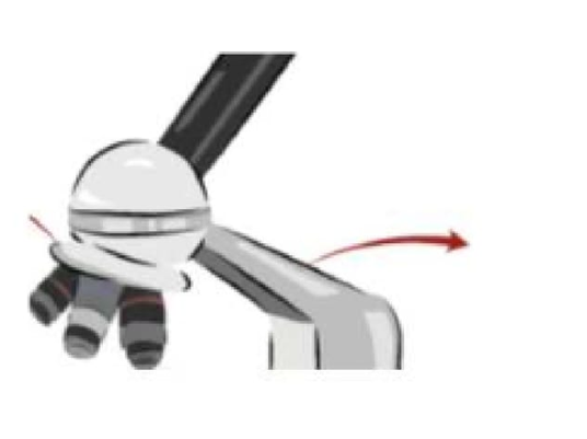
Arm
Shoulder
The Correct Answer is A
Based on the image provided, the arrow is pointing to the arm of the microscope.
Arm: The arm is the curved or slanted part of the microscope that connects the base to the head (which contains the eyepiece and revolving nosepiece). It serves two main purposes: It holds the upper portion of the microscope (the optical components) securely above the stage. It is the primary part of the microscope designed to be gripped when carrying the instrument.
Shoulder: The shoulder is the upper supporting part of the microscope where the arm meets the head or body tube. It provides structural support for the optical components and helps maintain proper alignment of the microscope parts.
Nursing Test Bank
Naxlex Comprehensive Predictor Exams
Related Questions
Correct Answer is A
Explanation
A. Loop & needle: In microbiology, a sterile inoculating loop or needle is commonly used to transfer microorganisms from one culture medium to another while maintaining aseptic technique. Loops are typically used for streaking or spreading bacteria on agar, while needles are used for stabbing into agar or transferring small amounts of culture into broth, allowing precise and sterile handling.
B. Loop & syringe: Syringes are generally not used for routine transfer of microbial cultures between media because they increase the risk of contamination and are more appropriate for measuring or injecting liquids rather than streaking or inoculating media.
C. Needle & syringe: Needles with syringes are mainly used in clinical or laboratory procedures for injecting liquids or drawing samples. They are not standard instruments for sterile transfer between culture media in microbiology because they are less precise for streaking or isolating colonies.
D. all above are true: Only the loop and needle are routinely used for sterile transfers in microbiology. Syringes are not standard instruments for transferring cultures between media, so this option is incorrect.
Correct Answer is B
Explanation
A. To stain the bacteria: Heat fixing does not apply stain to the bacteria; staining is a separate step performed after the smear is fixed. Heat fixing prepares the cells to better accept the stain by immobilizing them and preserving their structure.
B. To kill bacteria and adhere them to the slide: Heat fixing serves two main purposes: it kills the bacteria, making the slide safe to handle, and it causes the proteins in the cells to coagulate slightly, which adheres the cells firmly to the glass slide. This prevents them from washing off during the staining and rinsing and preserves their morphology for accurate examination.
C. To increase magnification: Heat fixing has no effect on the magnification of the microscope. Magnification is controlled by the objective lenses and ocular lenses, not by the preparation technique of the smear.
D. To remove excess stain: Heat fixing does not remove stain; rather, it prepares the cells so that stains can bind effectively. Removal of excess stain is achieved through rinsing with water or appropriate solvents after staining, not through heat fixation.
Whether you are a student looking to ace your exams or a practicing nurse seeking to enhance your expertise , our nursing education contents will empower you with the confidence and competence to make a difference in the lives of patients and become a respected leader in the healthcare field.
Visit Naxlex, invest in your future and unlock endless possibilities with our unparalleled nursing education contents today