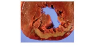
The left ventricle of the heart from a 70-year-old man is examined at autopsy (shown). What was the most likely cause of the patient's death?
Cardiac tamponade
Congestive heart failure
Hypovolemic shock
Myocardial infarction
Pulmonary thromboembolism
The Correct Answer is D
A. Cardiac tamponade: Cardiac tamponade occurs when fluid accumulates in the pericardial sac, compressing the heart and reducing cardiac output. Gross pathology shows pericardial effusion rather than a localized area of myocardial necrosis, making it an unlikely cause here.
B. Congestive heart failure: Congestive heart failure results from chronic ventricular dysfunction and presents with pulmonary congestion, peripheral edema, and hepatomegaly. It does not produce a well-demarcated, yellow-tan area of necrosis in the ventricular wall.
C. Hypovolemic shock: Hypovolemic shock is due to severe blood or fluid loss, leading to systemic hypotension and multi-organ hypoperfusion. The heart itself does not show a focal infarct as seen in this autopsy finding.
D. Myocardial infarction: A myocardial infarction occurs when coronary artery obstruction causes ischemic necrosis of the myocardium. The gross pathological finding of a well-defined yellow-tan area in the left ventricular wall corresponds to coagulative necrosis following MI and is the most likely cause of death in this patient.
E. Pulmonary thromboembolism: Pulmonary thromboembolism involves obstruction of pulmonary arteries by thrombus, which can cause acute right heart strain and sudden death. It does not produce a focal infarct in the left ventricular wall.
Nursing Test Bank
Naxlex Comprehensive Predictor Exams
Related Questions
Correct Answer is D
Explanation
A. Basophils:Basophils are circulating granulocytes involved in allergic reactions and parasitic infections. They release histamine and other mediators but do not form multinucleated giant cells or participate directly in granuloma formation in the lungs.
B. Endothelial cells:Endothelial cells line blood vessels and are involved in vascular permeability and inflammation. They do not differentiate into multinucleated giant cells and are not a source of granulomatous structures.
C. Eosinophils:Eosinophils are primarily involved in parasitic infections and allergic responses. While they may be present in some inflammatory infiltrates, they do not fuse to form the multinucleated giant cells seen in granulomatous lung disease.
D. Macrophages:Multinucleated giant cells in granulomas are derived from activated macrophages. These macrophages fuse in response to persistent antigens, such as Mycobacterium tuberculosisor fungi, and coordinate a chronic inflammatory response to contain pathogens that are difficult to eradicate.
E. Neutrophils:Neutrophils are the first responders in acute inflammation and are effective in phagocytosing bacteria. They do not fuse to form multinucleated giant cells, which are a hallmark of chronic granulomatous inflammation.
Correct Answer is E
Explanation
A. Bacterial pneumonia:Bacterial pneumonia can cause dyspnea and rales, but it typically presents with fever, localized infiltrates on imaging, and productive cough. In this patient, the acute onset following extensive burns points toward non-infectious pulmonary injury rather than a primary bacterial infection.
B. Cardiogenic shock:Cardiogenic shock results from acute cardiac pump failure, leading to hypotension and poor organ perfusion. While pulmonary edema may accompany cardiogenic shock, there is no indication in this scenario that the patient has primary cardiac dysfunction; the burns and systemic inflammatory response are the key factors.
C. Congestive heart failure:Congestive heart failure causes pulmonary congestion and rales due to left ventricular dysfunction. This patient’s diffuse alveolar damage is a direct result of burn-induced systemic inflammation rather than chronic or acute cardiac failure, making CHF less likely.
D. Dehydration (hypovolemic shock):Hypovolemic shock from fluid loss in burns leads to low blood pressure and poor tissue perfusion. It does not cause pulmonary edema or alveolar rales, which are related to fluid accumulation in the lungs rather than intravascular volume depletion.
E. Pulmonary edema:Diffuse alveolar damage from burn injuries increases alveolar-capillary permeability, allowing protein-rich fluid to accumulate in alveoli. This results in pulmonary edema, causing rales on auscultation and dyspnea due to impaired gas exchange, making it the most likely cause of the patient’s respiratory findings.
Whether you are a student looking to ace your exams or a practicing nurse seeking to enhance your expertise , our nursing education contents will empower you with the confidence and competence to make a difference in the lives of patients and become a respected leader in the healthcare field.
Visit Naxlex, invest in your future and unlock endless possibilities with our unparalleled nursing education contents today