The plate shown below is known as:
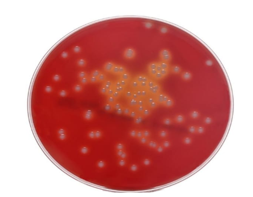
Blood agar plate (BAP)
M17 Agar
The Correct Answer is A
A blood agar plate is a nutrient-rich culture medium that contains mammalian blood, usually 5% sheep blood, mixed with agar. It is commonly used in microbiology laboratories to grow and differentiate bacteria based on their ability to lyse red blood cells (hemolysis). The red color of the medium comes from the intact red blood cells, and bacterial colonies may produce clear zones (beta hemolysis), greenish zones (alpha hemolysis), or no change (gamma hemolysis) around them. This property helps in identifying clinically important bacteria such as Streptococcus species.
Nursing Test Bank
Naxlex Comprehensive Predictor Exams
Related Questions
Correct Answer is A
Explanation
Gamma hemolysis, also called non-hemolysis, refers to bacterial growth on blood agar that does not cause any breakdown of red blood cells. On a blood agar plate, colonies exhibiting gamma hemolysis appear unchanged, with no clear or greenish zones surrounding them, indicating the bacteria do not produce hemolysins that lyse erythrocytes. This contrasts with alpha hemolysis, which causes partial hemolysis and greenish discoloration, and beta hemolysis, which produces complete lysis and clear zones around colonies. Therefore, gamma hemolysis signifies the absence of red blood cell lysis.
Correct Answer is C
Explanation
A. Purple: Purple indicates Gram-positive bacteria, which retain the crystal violet-iodine complex within their thick peptidoglycan cell walls during the Gram staining process. The color persists even after decolorization with alcohol or acetone.
B. Blue: Blue is not a standard color outcome in Gram staining. Gram-negative and Gram-positive bacteria are typically differentiated as purple or pink/red; blue may appear in other specialized staining techniques but not standard Gram staining.
C. Pink/Red: Gram-negative bacteria appear pink or red after Gram staining. Their thin peptidoglycan layer does not retain the crystal violet-iodine complex after decolorization. They are counterstained with safranin, which imparts the pink/red color, allowing differentiation from Gram-positive bacteria.
D. Green: Green is not a result of Gram staining. Green coloration is associated with other staining methods or pigments but is not part of the Gram stain differential process.
Whether you are a student looking to ace your exams or a practicing nurse seeking to enhance your expertise , our nursing education contents will empower you with the confidence and competence to make a difference in the lives of patients and become a respected leader in the healthcare field.
Visit Naxlex, invest in your future and unlock endless possibilities with our unparalleled nursing education contents today