The trigone of the bladder is formed by the two ureteric orifices and the internal urethral opening.
True
False
The Correct Answer is A
A. True: The trigone is a smooth, triangular region located at the base of the urinary bladder. Its boundaries are precisely defined by the entry points of both ureters and the exit point of the urethra. This area is clinically significant and remains fixed during bladder expansion.
Nursing Test Bank
Naxlex Comprehensive Predictor Exams
Related Questions
Correct Answer is C
Explanation
A. It causes water to move out of cells, shrinking them: This occurs in a hyper-osmolar environment, where the high concentration of extracellular solutes draws water out via osmosis. Hypo-osmolarity involves a lower solute concentration outside the cell. The osmotic pressure gradient would favor influx, not efflux.
B. It increases vascular resistance, raising blood pressure: Hypo-osmolarity is often associated with fluid overload, but it does not directly cause vasoconstriction. In many cases, it is linked to low sodium levels which can impair vascular tone. It primarily affects fluid distribution rather than active vessel resistance.
C. It causes water to move into cells, swelling and possible rupture: In a hypo-osmolar state, the intracellular fluid has a higher solute concentration than the surrounding plasma. Water moves down its osmotic gradient into the cells to achieve equilibrium. This cellular edema can lead to lysis and organ dysfunction.
D. It enhances sodium retention, preventing edema: Hypo-osmolarity typically triggers mechanisms to excrete water or retain sodium to restore balance. However, if the condition persists, the low osmotic pressure in the blood allows fluid to leak into the interstitial spaces. This results in the formation of edema.
Correct Answer is {"dropdown-group-1":"D","dropdown-group-2":"E","dropdown-group-3":"E"}
Explanation
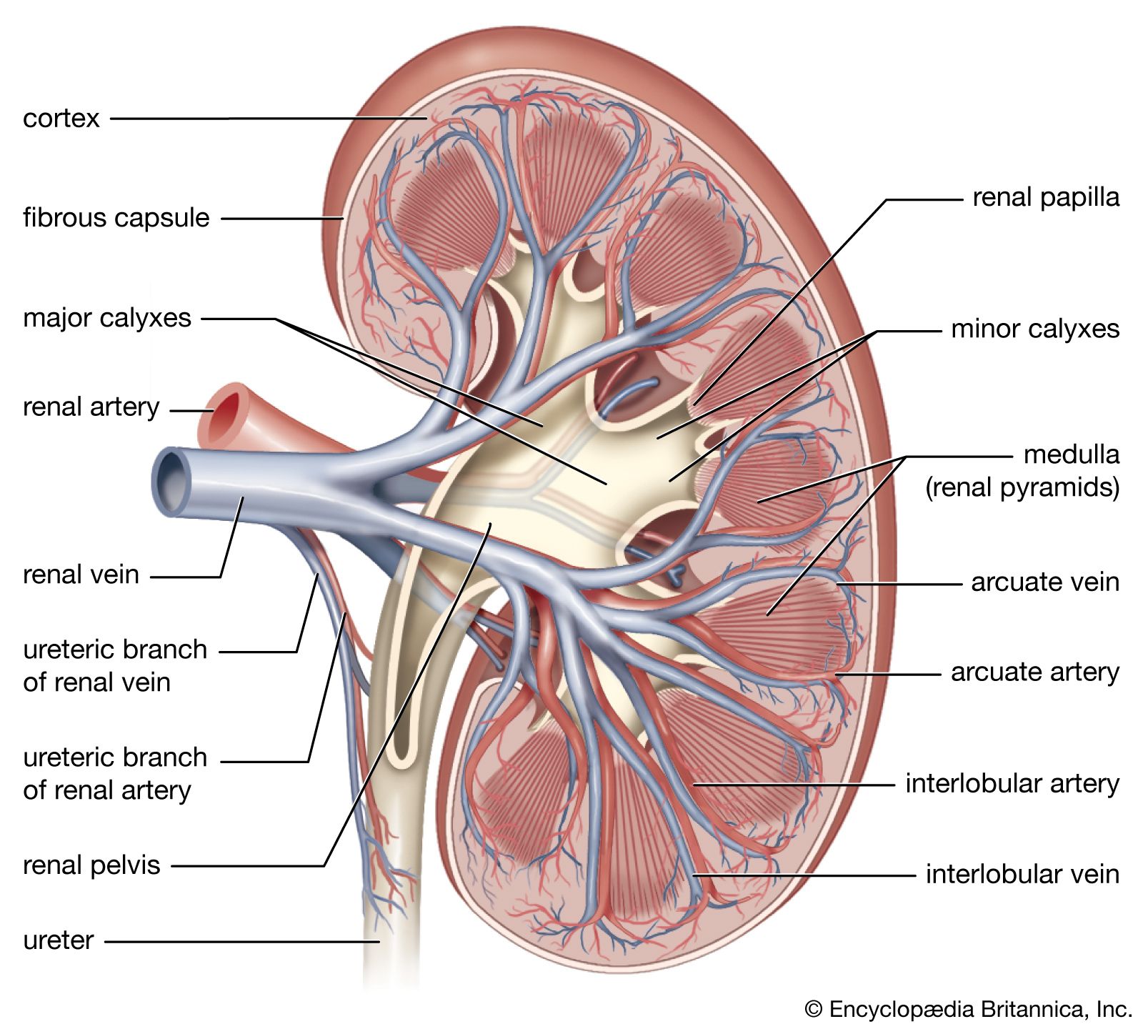
Minor calyx (I): The minor calyx is a small, cup-shaped structure that collects urine from the renal papilla at the tip of each pyramid. It is part of the drainage system, channeling urine into the major calyces and then into the renal pelvis. Look for the small funnel-like extensions directly attached to the renal papillae. These are distinctly smaller than the major calyces.
Renal medulla(H): The renal medulla is the inner region of the kidney, composed of renal pyramids. It plays a crucial role in concentrating urine through the countercurrent mechanism in the loops of Henle and collecting ducts. Appears as darker, triangular or striated regions deeper inside the kidney, between the cortex and the renal pelvis.
Renal cortex (F): The renal cortex is the outer functional tissue of the kidney, lying just beneath the fibrous capsule. It contains glomeruli and convoluted tubules, making it the primary site of blood filtration. Appears as the lighter-colored outer rim of the kidney, surrounding the medulla.
Whether you are a student looking to ace your exams or a practicing nurse seeking to enhance your expertise , our nursing education contents will empower you with the confidence and competence to make a difference in the lives of patients and become a respected leader in the healthcare field.
Visit Naxlex, invest in your future and unlock endless possibilities with our unparalleled nursing education contents today