Use the diagram above to answer the following questions.
Identify the letter that indicates the cardia of the stomach.
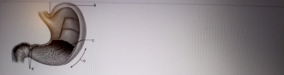
A
B
C
D
E
The Correct Answer is A
A. Cardia: This is the narrow, superior region of the stomach located immediately distal to the gastroesophageal junction where the esophagus enters the gastric chamber. It contains the cardiac glands, which primarily secrete mucus to protect the esophageal lining from the highly acidic environment of the stomach.
B. Fundus: This is the dome-shaped, superior-most portion of the stomach that sits tucked under the left dome of the diaphragm, often accumulating swallowed air. It serves as a temporary storage area for undigested food and contains a high density of gastric glands that produce acid and digestive enzymes.
C. Body/Corpus: This represents the large, central region of the stomach between the fundus and the pyloric antrum, serving as the main mixing tank for ingested food. The rugae muscular walls in this area undergo rhythmic peristaltic contractions to mechanically break down food into a semi-liquid substance known as chyme.
D. Greater Curvature: This is the convex, lateral border of the stomach that provides an attachment point for the greater omentum, a significant fold of the peritoneum. It is much longer than the medial lesser curvature and marks the outer boundary of the gastric body and fundus.
E. Pyloric Sphincter: The pyloric sphincter is a thick, circular band of smooth muscle located at the distal end of the stomach, where the stomach connects to the duodenum. It acts as a gatekeeper, regulating the release of chyme into the duodenum to ensure that the small intestine is not overwhelmed by large volumes of acidic contents.
Nursing Test Bank
Naxlex Comprehensive Predictor Exams
Related Questions
Correct Answer is A
Explanation
A. Sternocleidomastoid muscle: This muscle arises from the manubrium of the sternum and the clavicle, inserting onto the mastoid process of the temporal bone. It mainly facilitates head rotation to the opposite side and neck flexion, and serves as a key landmark in the anterior neck region.
B. Deltoid muscle: The deltoid is a thick, multipennate muscle forming the rounded shape of the shoulder. It originates from the lateral third of the clavicle, acromion, and spine of the scapula, and its primary role is abducting the arm away from the body’s midline.
C. Pectoralis major: This thick, fan-shaped muscle, makes up the bulk of the chest muscles and is responsible for adduction and internal rotation of the humerus. It originates from the clavicle, sternum, and costal cartilages. It plays a significant role in powerful upper limb movements such as pushing, throwing, or climbing.
D. Pectoralis minor: This thin, triangular muscle sits deep to the pectoralis major and originates from the third, fourth, and fifth ribs. Its primary function is to stabilize the scapula by drawing it inferiorly and anteriorly against the thoracic wall.
E. Serratus anterior: The serratus anterior originates on the surface of the upper eight or nine ribs and inserts along the entire anterior length of the medial border of the scapula. It is often referred to as the "boxer's muscle" because it is essential for the protraction of the scapula, such as when throwing a punch.
Correct Answer is D
Explanation
A. Neutrophil fraction: The neutrophil fraction, which is the most abundant type of leukocyte, typically accounts for 50% to 70% of the total white blood cell count. These granulocytes are the primary responders to acute bacterial infections and utilize phagocytosis and degranulation as their main effector mechanisms, making them the largest component of the differential count.
B. Eosinophil fraction: This type of granulocyte that usually accounts for only 2% to 4% of the total white blood cell population. These cells are specialized for the defense against multicellular parasites and are significant mediators of allergic inflammatory responses, but they represent a much smaller percentage of the differential than lymphocytes.
C. Basophil fraction: The basophil fraction is the rarest leukocyte type, normally constituting less than 1% of the total white blood cell count. Basophils contain large granules filled with histamine and heparin and are primarily involved in systemic inflammatory and hypersensitivity reactions, making them the smallest identifiable segment in a standard leukocyte differential.
D. Lymphocyte fraction: The lymphocyte fraction accounts for approximately 20% to 40% of the total count. Lymphocytes, including B-cells, T-cells, and Natural Killer (NK) cells, are the central mediators of adaptive immunity and are responsible for antigen recognition, antibody production, and the destruction of virally infected cells.
E. Monocyte fraction: The monocyte fraction makes up about 3% to 8% of the circulating leukocytes and represents the largest individual cell type in the blood. Monocytes are agranulocytes that migrate into tissues to differentiate into macrophages and dendritic cells, serving as critical antigen-presenting cells that link the innate and adaptive immune responses.
Whether you are a student looking to ace your exams or a practicing nurse seeking to enhance your expertise , our nursing education contents will empower you with the confidence and competence to make a difference in the lives of patients and become a respected leader in the healthcare field.
Visit Naxlex, invest in your future and unlock endless possibilities with our unparalleled nursing education contents today