Use the diagrams above to answer the following questions.
Identify which letter represents an oligodendrocyte.
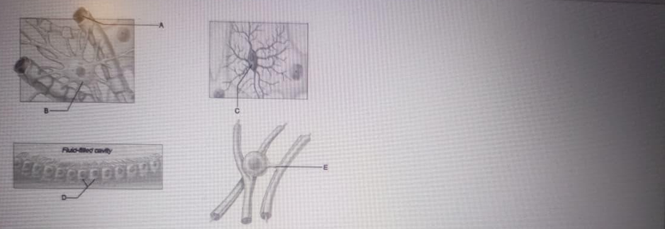
A
B
C
D
E
The Correct Answer is E
A. Astrocyte: This is the most abundant neuroglial cell in the central nervous system, characterized by its star-shaped morphology and perivascular feet. These cells are essential for maintaining the blood-brain barrier, regulating the chemical environment of the interstitial fluid, and providing structural support to neurons. Their primary role is homeostatic and metabolic.
B. Schwann Cells: Schwann cells are glial cells of the peripheral nervous system that form the myelin sheath around axons. This myelination increases the speed of electrical impulse conduction along the nerve fiber. Schwann cells also aid in the regeneration of damaged peripheral nerves by guiding axonal growth.
C. Microglial cell: This cell functions as the resident macrophage and primary immune defense within the central nervous system. These small, mobile cells constantly scavenge for plaque, damaged neurons, and infectious agents to maintain neural health through phagocytosis.
D. Ependymal cells: Ependymal cells line the ventricles of the brain and the central canal of the spinal cord. These ciliated epithelial-like cells are responsible for the production, circulation, and monitoring of cerebrospinal fluid (CSF), creating a permeable barrier between the CSF and the nervous tissue. Their specialization is related to fluid dynamics and ventricular lining.
E. Oligodendrocyte: This is a specialized glial cell that extends multiple cytoplasmic processes to wrap around axons in the central nervous system. These wraps form the myelin sheath, a lipid-rich insulating layer that significantly increases the velocity of action potential conduction through saltatory conduction.
Nursing Test Bank
Naxlex Comprehensive Predictor Exams
Related Questions
Correct Answer is A
Explanation
A. Cardia: This is the narrow, superior region of the stomach located immediately distal to the gastroesophageal junction where the esophagus enters the gastric chamber. It contains the cardiac glands, which primarily secrete mucus to protect the esophageal lining from the highly acidic environment of the stomach.
B. Fundus: This is the dome-shaped, superior-most portion of the stomach that sits tucked under the left dome of the diaphragm, often accumulating swallowed air. It serves as a temporary storage area for undigested food and contains a high density of gastric glands that produce acid and digestive enzymes.
C. Body/Corpus: This represents the large, central region of the stomach between the fundus and the pyloric antrum, serving as the main mixing tank for ingested food. The rugae muscular walls in this area undergo rhythmic peristaltic contractions to mechanically break down food into a semi-liquid substance known as chyme.
D. Greater Curvature: This is the convex, lateral border of the stomach that provides an attachment point for the greater omentum, a significant fold of the peritoneum. It is much longer than the medial lesser curvature and marks the outer boundary of the gastric body and fundus.
E. Pyloric Sphincter: The pyloric sphincter is a thick, circular band of smooth muscle located at the distal end of the stomach, where the stomach connects to the duodenum. It acts as a gatekeeper, regulating the release of chyme into the duodenum to ensure that the small intestine is not overwhelmed by large volumes of acidic contents.
Correct Answer is E
Explanation
A. Cervical sympathetic trunk: This is the superior portion of the sympathetic trunk in the cervical or upper thoracic region, far from the pelvic cavity. It provides sympathetic innervation to the head, neck, and upper thoracic viscera, such as the heart and lungs, through the cervical and superior thoracic paravertebral ganglia.
B. Thoracic sympathetic trunk: The thoracic sympathetic trunk runs parallel to the vertebral column within the chest cavity and gives rise to the splanchnic nerves. These nerves primarily target the prevertebral ganglia in the abdomen to regulate the function of the foregut and midgut, rather than descending into the true pelvis.
C. Lumbar sympathetic trunk: The lumbar sympathetic trunk is situated along the lumbar vertebrae and responsible for providing postganglionic fibers to the lower abdominal region and lower extremities. While it is continuous with the pelvic portion, it is anatomically distinct and superior to the sacral/pelvic region of the autonomic chain.
D. Abdominal prevertebral plexus: This points to the abdominal prevertebral plexus (specifically near the aortic bifurcation), which integrates both sympathetic and parasympathetic fibers. While these plexuses contribute to the pelvic viscera, they are located anterior to the great vessels and do not constitute the paravertebral "trunk" or chain itself.
E. Pelvic (sacral) sympathetic trunk: This identifies the pelvic (sacral) sympathetic trunk, which consists of the paravertebral ganglia located medial to the sacral foramina. These ganglia represent the terminal inferior portion of the sympathetic chain, where the two trunks converge at the coccyx to form the ganglion impar, providing sympathetic outflow to the pelvic organs and perineum.
Whether you are a student looking to ace your exams or a practicing nurse seeking to enhance your expertise , our nursing education contents will empower you with the confidence and competence to make a difference in the lives of patients and become a respected leader in the healthcare field.
Visit Naxlex, invest in your future and unlock endless possibilities with our unparalleled nursing education contents today