Which letter on the image depicts the QT segment?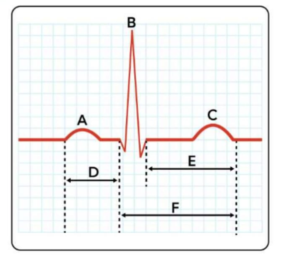
D
A
C
F
The Correct Answer is D
A. P wave:: The P wave represents atrial depolarization, indicating electrical activation of the atria. It precedes atrial contraction and is the first deflection on a standard ECG tracing. Normal duration is ≤0.12 seconds.
C. T wave: The T wave reflects ventricular repolarization, showing the recovery phase of the ventricles after contraction. Its shape and amplitude can indicate electrolyte imbalances, ischemia, or other cardiac abnormalities.
F. QT segment: The QT segment (more accurately called the QT interval), labelled as F, represents the total time for ventricular depolarization and repolarization. It begins at the start of the QRS complex and ends at the conclusion of the T wave on an electrocardiogram (ECG). It reflects the entire period of ventricular electrical activity, including contraction and recovery.
D. PR interval: The PR interval represents the time for electrical conduction from the atria through the AV node to the ventricles. Normal duration is 0.12–0.20 seconds, with prolongation suggesting conduction delay.
Nursing Test Bank
Naxlex Comprehensive Predictor Exams
Related Questions
Correct Answer is C
Explanation
A. Myoglobin and lipid inclusions: Both skeletal and cardiac muscle fibers contain myoglobin, which stores oxygen for aerobic metabolism, and lipid inclusions as energy reserves. These features support high metabolic demands in both muscle types and are not unique to skeletal muscle.
B. A single nucleus per cell: Cardiac muscle cells are typically uninucleated, whereas skeletal muscle fibers are multinucleated. However, the presence of a single nucleus is characteristic of cardiac myocytes, not a distinguishing feature of skeletal muscle.
C. Triads formed by long T tubules and cisternae of the sarcoplasmic reticulum: Skeletal muscle fibers have well-organized triads, where a T tubule is flanked by two terminal cisternae of the sarcoplasmic reticulum, allowing rapid calcium release for synchronous contraction. In contrast, cardiac contractile cells have diads (one T tubule with one adjacent cisterna) and smaller T tubules, reflecting slower calcium handling.
D. Sarcomeres along myofibrils: Both skeletal and cardiac muscle fibers contain sarcomeres arranged along myofibrils, giving them striated appearance under microscopy. Sarcomeres are essential for contraction in both types of striated muscle and are not a distinguishing structural feature.
Correct Answer is {"dropdown-group-1":"C","dropdown-group-2":"B"}
Explanation
Correct answer:
- Interventricular septum
- Atrioventricular (AV) bundle / Bundle of His
The highlighted area represents the interventricular septum, the thick muscular wall that separates the right and left ventricles. It forms the medial wall of both ventricles and extends from the atrioventricular valves superiorly to the apex inferiorly. Its primary physiologic function is to prevent mixing of oxygenated blood in the left ventricle with deoxygenated blood in the right ventricle while also contributing to ventricular contraction. The electrical conduction structure that passes through this area is the atrioventricular (AV) bundle / Bundle of His. The Bundle of His passes from the atrioventricular node into the membranous portion of the interventricular septum. It then divides into the right and left bundle branches, which travel along the septum toward the apex to distribute electrical impulses to both ventricles. This allows coordinated ventricular depolarization and synchronized contraction.
Whether you are a student looking to ace your exams or a practicing nurse seeking to enhance your expertise , our nursing education contents will empower you with the confidence and competence to make a difference in the lives of patients and become a respected leader in the healthcare field.
Visit Naxlex, invest in your future and unlock endless possibilities with our unparalleled nursing education contents today