Which structure is indicated by the highlight and leader line?
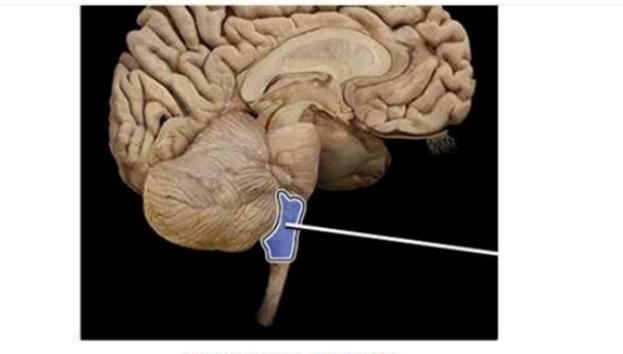
Pons
Medulla oblongata
Midbrain
Cerebellum
The Correct Answer is B
The marked structure is the medulla oblongata, the most inferior portion of the brainstem, continuous with the spinal cord at the foramen magnum. It lies between the pons superiorly and the spinal cord inferiorly and contains both ascending and descending neural tracts. The medulla plays a vital role in autonomic control centers regulating respiration, heart rate, and blood pressure. It also contains nuclei of several cranial nerves (IX–XII), making it essential for life-sustaining reflexes.
A. Pons: The pons is the middle segment of the brainstem located superior to the medulla and inferior to the midbrain. It serves as a bridge (“pons” meaning bridge) connecting the cerebrum with the cerebellum via transverse pontine fibers. It also plays a role in regulating respiration and relaying motor and sensory information. Compared to the medulla, it is more rounded and positioned superiorly in the brainstem.
B. Medulla oblongata: The medulla oblongata is the most caudal part of the brainstem, continuous with the spinal cord. It contains vital autonomic centers controlling respiration, cardiac rhythm, and vasomotor tone. It houses nuclei for cranial nerves IX, X, XI, and XII and coordinates reflexes such as swallowing, coughing, and vomiting. Its location just above the spinal cord and below the pons makes it the correct answer.
C. Midbrain: The midbrain is the superior portion of the brainstem located between the pons and the diencephalon. It is involved in visual and auditory reflexes and contains structures such as the superior and inferior colliculi. It is more rostral and smaller compared to the medulla and does not extend into the spinal cord region.
D. Cerebellum: The cerebellum is located posterior to the brainstem in the posterior cranial fossa. It is responsible for coordination of voluntary movement, balance, posture, and motor learning. Unlike the medulla, it is not part of the brainstem’s autonomic control system and does not regulate vital life-sustaining functions like respiration or cardiac output.
Nursing Test Bank
Naxlex Comprehensive Predictor Exams
Related Questions
Correct Answer is D
Explanation
Sensory receptors are specialized structures that detect different types of stimuli and transmit information to the central nervous system for processing. They are classified based on the type of stimulus they detect, including pressure, light, pain, and body position. In movement and coordination, certain receptors provide continuous feedback about body position in space. This is essential for balance, posture, and coordinated athletic performance such as in activities requiring mid-air awareness.
A. Baroreceptors: Baroreceptors are mechanoreceptors located primarily in the carotid sinus and aortic arch. They detect changes in blood pressure by sensing stretch in the arterial walls. When blood pressure rises or falls, they send signals to the medulla to regulate heart rate and vascular tone. They do not provide information about limb position or spatial awareness during movement, making them unrelated to mid-air body tracking.
B. Photoreceptors: Photoreceptors are specialized sensory cells located in the retina of the eye, consisting of rods and cones. Rods detect low-light conditions, while cones are responsible for color vision and visual acuity. Their function is to convert light energy into electrical signals for visual perception. Although they contribute to spatial awareness through vision, they do not directly provide internal feedback about body position in space.
C. Nociceptors: Nociceptors are pain receptors found in skin, muscles, joints, and internal organs. They respond to potentially damaging stimuli such as extreme temperature, mechanical injury, or chemical irritation. Their primary function is to initiate pain perception as a protective mechanism. While they help detect injury, they do not provide information about body position or movement coordination in space.
D. Proprioceptors: Proprioceptors are specialized mechanoreceptors located in muscles, tendons, and joint capsules, including muscle spindles and Golgi tendon organs. They continuously monitor muscle length, tension, and joint position, sending this information to the central nervous system. This allows the brain to maintain awareness of body position, coordination, and balance without visual input. In activities like pole vaulting, proprioceptors enable precise mid-air spatial orientation and controlled landing.
Correct Answer is B
Explanation
Skeletal muscle fibers are highly specialized cells designed for rapid and coordinated contraction. To achieve this, they require an efficient system for transmitting electrical signals from the cell surface deep into the muscle fiber. Transverse (T) tubules are invaginations of the sarcolemma that penetrate into the cell interior. They work closely with the sarcoplasmic reticulum to ensure uniform and rapid activation of muscle contraction throughout the fiber.
A. To store calcium ions needed for activating tropomyosin: calcium storage in muscle cells is primarily handled by the sarcoplasmic reticulum, not the T-tubules. The sarcoplasmic reticulum releases calcium ions in response to an action potential, allowing calcium to bind troponin and shift tropomyosin away from actin binding sites. T-tubules do not store calcium; they serve as conduits for electrical signals.
B. To transmit action potentials (impulses) into the cell interior: transverse tubules are invaginations of the sarcolemma that rapidly conduct action potentials from the cell surface into the deeper regions of the muscle fiber. This ensures that the entire muscle fiber contracts simultaneously rather than in a wave-like fashion. The electrical signal traveling through T-tubules triggers calcium release from the sarcoplasmic reticulum. This coupling is essential for coordinated and efficient muscle contraction.
C. To synthesize ATP for muscle contraction: ATP production occurs primarily in mitochondria through oxidative phosphorylation and in the cytoplasm via glycolysis. T-tubules have no role in energy metabolism or ATP synthesis. Their function is electrical signal transmission, not biochemical energy production. Therefore, this option describes a mitochondrial function rather than a T-tubule function.
D. To break down acetylcholine at the neuromuscular junction: acetylcholine breakdown is performed by the enzyme acetylcholinesterase located in the synaptic cleft of the neuromuscular junction. T-tubules are located inside the muscle fiber and are not involved in synaptic transmission or neurotransmitter degradation.
Whether you are a student looking to ace your exams or a practicing nurse seeking to enhance your expertise , our nursing education contents will empower you with the confidence and competence to make a difference in the lives of patients and become a respected leader in the healthcare field.
Visit Naxlex, invest in your future and unlock endless possibilities with our unparalleled nursing education contents today