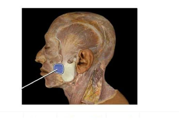
Which structure is highlighted in the image below?
Masseter
Zygomatic
Buccinator
Temporalis
The Correct Answer is A
The marked structure is the masseter muscle, one of the primary muscles of mastication located on the lateral aspect of the mandible. It originates from the zygomatic arch and inserts onto the lateral surface of the ramus and angle of the mandible. The masseter is one of the strongest muscles in the body relative to its size and is essential for forceful elevation of the mandible during chewing. It works in coordination with the temporalis and pterygoid muscles to produce efficient grinding and crushing of food during mastication.
A. Masseter: The masseter is a thick, rectangular muscle situated over the lateral surface of the mandibular ramus. It elevates the mandible, producing powerful jaw closure required for chewing tough food. It has superficial and deep layers and is easily visible when the jaw is clenched. Its location over the angle of the jaw and strong vertical fibers make it the correct structure.
B. Zygomatic: The zygomatic region refers to the zygomatic bone (cheekbone), which forms the prominence of the cheek and part of the orbital rim. It is a bone, not a muscle, and serves as an attachment site for facial muscles. Unlike the masseter, it does not contract or contribute to jaw movement.
C. Buccinator: The buccinator is a thin, flat muscle located in the cheek. It assists in compressing the cheek against the teeth, aiding in chewing by keeping food between the occlusal surfaces. It is also involved in blowing and whistling. Unlike the masseter, it is deep and does not produce strong jaw elevation.
D. Temporalis: The temporalis is a fan-shaped muscle located on the lateral skull in the temporal fossa. It elevates and retracts the mandible and plays a key role in closing the jaw. Although it is also a muscle of mastication, it is positioned superiorly on the skull rather than over the lateral jaw like the masseter.
Nursing Test Bank
Naxlex Comprehensive Predictor Exams
Related Questions
Correct Answer is D
Explanation
The ear is divided into the outer, middle, and inner ear, each contributing to the process of hearing and balance. The middle ear contains a chain of three small bones known as the auditory ossicles. These structures play a crucial role in transmitting sound vibrations from the tympanic membrane to the inner ear. Their arrangement allows mechanical amplification of sound and efficient transfer of energy into the fluid-filled cochlea for auditory processing.
A. Receptors for hearing, static equilibrium, and dynamic equilibrium: the malleus, incus, and stapes are not sensory receptors. Hearing and balance receptors are located in the inner ear, specifically within the cochlea and vestibular apparatus. Hair cells in the organ of Corti detect sound, while hair cells in the semicircular canals, utricle, and saccule detect equilibrium. The ossicles instead serve a mechanical role in transmitting vibrations.
B. Regions of the inner ear: the ossicles are not part of the inner ear. The inner ear includes structures such as the cochlea, vestibule, and semicircular canals, which are responsible for hearing and balance. The malleus, incus, and stapes are located in the middle ear cavity between the tympanic membrane and oval window. They function externally to the inner ear structures rather than within them.
C. Types of mechanoreceptors: the ossicles are bones, not sensory receptors. Mechanoreceptors are specialized nerve endings that detect mechanical stimuli such as pressure, stretch, and vibration. The malleus, incus, and stapes do not transduce sensory information but instead physically transmit sound vibrations. Therefore, they cannot be classified as mechanoreceptors.
D. Bones of the middle ear: the malleus, incus, and stapes are the three auditory ossicles located in the middle ear. The malleus attaches to the tympanic membrane, the incus serves as the connecting bone, and the stapes interfaces with the oval window of the cochlea. Together, they form a mechanical linkage that amplifies and transmits sound vibrations from air to fluid media, enabling efficient hearing.
Correct Answer is A
Explanation
The marked structure is the humerus, which is the long bone of the upper arm extending from the shoulder to the elbow joint. It articulates proximally with the scapula at the glenohumeral joint and distally with the radius and ulna at the elbow. The humerus plays a key role in upper limb movement, serving as an attachment site for multiple muscles involved in flexion, extension, rotation, and lifting. It is essential for both gross motor function and fine upper extremity coordination.
A. Humerus: The humerus is the long bone of the upper arm located between the shoulder and elbow joints. It articulates proximally with the scapula and distally with both the radius and ulna. It serves as the primary lever for upper limb movement and muscle attachment for the biceps, triceps, and deltoid.
B. Radius; The radius is one of the two forearm bones, located on the lateral (thumb) side of the forearm. It primarily articulates with the humerus at the elbow and with the carpal bones at the wrist, enabling pronation and supination. Unlike the humerus, it does not form the upper arm structure but instead functions in forearm rotation. This makes it anatomically distal to the marked upper arm bone.
C. Ulna: The ulna is the medial forearm bone, positioned on the side of the little finger. It forms the primary hinge joint with the humerus at the elbow, providing stability during flexion and extension. However, it does not extend into the upper arm region and is not involved in shoulder articulation. Compared to the humerus, it is a forearm stabilizing bone rather than the main upper limb shaft.
D. Femur: The femur is the long bone of the thigh and the strongest bone in the body, extending from the hip joint to the knee joint. It supports weight-bearing and locomotion in the lower extremity. Although it is also a long bone like the humerus, it is located in the lower limb and has no anatomical relationship to the upper arm region.
Whether you are a student looking to ace your exams or a practicing nurse seeking to enhance your expertise , our nursing education contents will empower you with the confidence and competence to make a difference in the lives of patients and become a respected leader in the healthcare field.
Visit Naxlex, invest in your future and unlock endless possibilities with our unparalleled nursing education contents today