A nurse is planning to assess the point of maximal impulse (PMI). What client position should the nurse use to enhance palpation of the PMI?
Prone with the arms overhead
Trendelenburg position
Supine and left lateral positions
Sitting upright with arms crossed
The Correct Answer is C
Choice A reason: The prone position is contraindicated for cardiac palpation because the client is lying face down, making the precordium inaccessible. This position is typically used for assessing the posterior lung fields or specific musculoskeletal issues but prevents the nurse from identifying the apical impulse located on the anterior-lateral aspect of the thoracic wall.
Choice B reason: The Trendelenburg position, where the head is lower than the feet, is used to promote venous return or during certain surgical procedures. It does not facilitate PMI palpation and may cause abdominal contents to push against the diaphragm, potentially displacing the heart superiorly and making the apical impulse more difficult to locate accurately.
Choice C reason: Palpating the PMI is best performed with the client supine, but if the impulse is difficult to feel, the left lateral decubitus position is utilized. This maneuver allows the apex of the heart to displace closer to the chest wall, making the pulsation of the left ventricle more palpable at the fifth intercostal space.
Choice D reason: While sitting upright is a standard position for auscultation, it is not the optimal position for enhancing the palpability of a faint PMI. Gravity in the seated position does not bring the cardiac apex forward as effectively as the left lateral position, which is the gold standard for magnifying the ventricular thrust.
Nursing Test Bank
Naxlex Comprehensive Predictor Exams
Related Questions
Correct Answer is B
Explanation
Choice A reason: Pain during chest expansion, often called pleuritic chest pain, is an abnormal finding. It may indicate pleurisy, where the pleural layers are inflamed and rub against each other. A healthy client should be able to take a deep breath comfortably without any stabbing or localized pain sensations during the maneuver.
Choice B reason: During the assessment of thoracic expansion, the nurse places their thumbs at the level of T9 or T10. As the client inhales deeply, the nurse's hands should move outward symmetrically. This indicates that both lungs are inflating equally and that the musculoskeletal structures of the chest wall are functioning uniformly on both sides.
Choice C reason: Asymmetrical rise and fall of the rib cage is a significant clinical abnormality. It suggests that one lung is not expanding fully, which can be seen in conditions such as massive atelectasis, pneumothorax, or pleural effusion on the affected side. This finding necessitates immediate further diagnostic investigation to determine the underlying cause of the restriction.
Choice D reason: A total lack of movement of the rib cage during a deep breath would indicate a profound respiratory failure, severe restrictive disease, or a neurological impairment of the diaphragm and intercostal muscles. In a living, breathing client, there should always be a palpable excursion of the thoracic cage during the inspiratory phase.
Correct Answer is C
Explanation
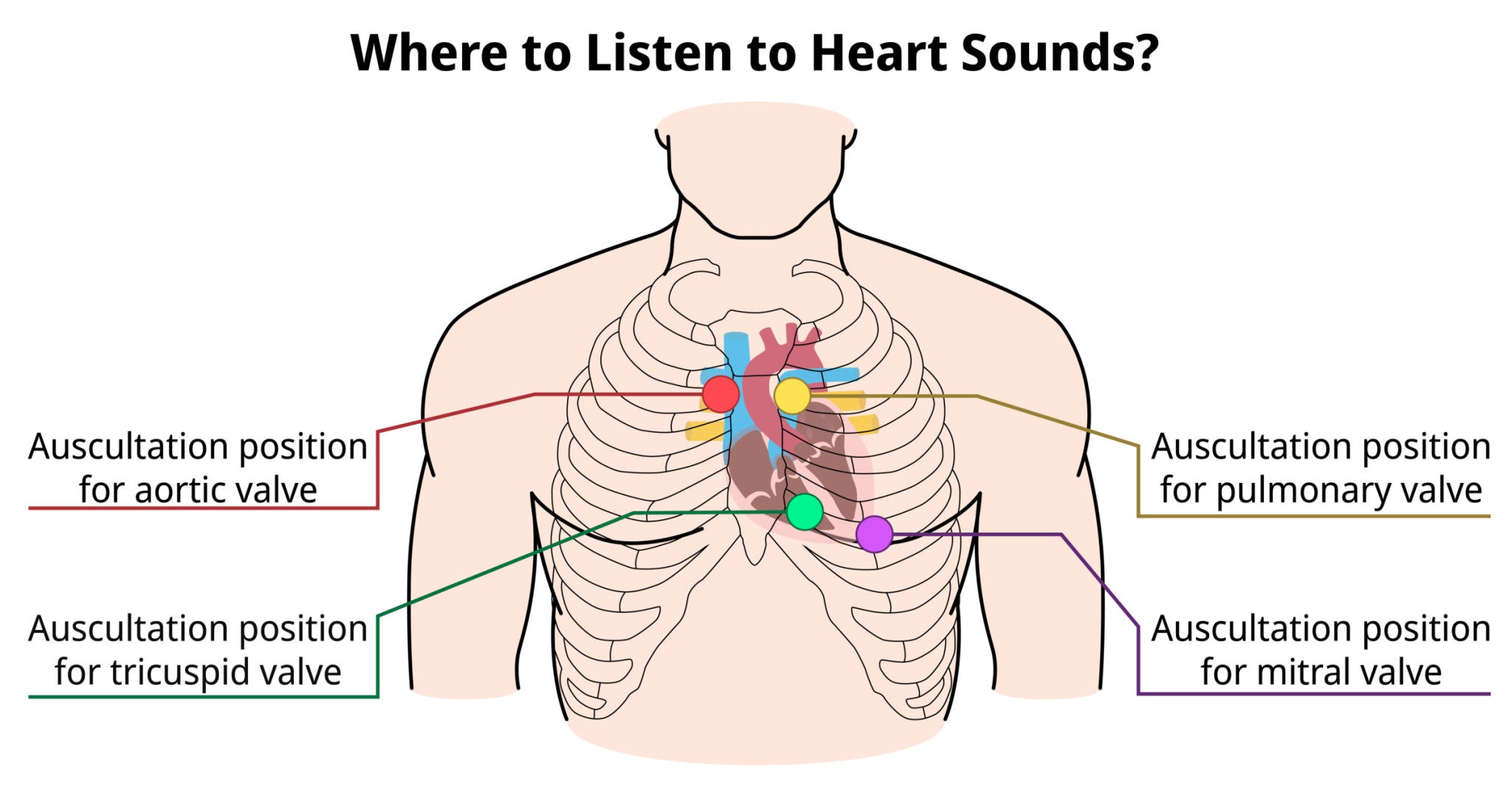
Choice A reason: The tricuspid valve is typically auscultated at the fourth or fifth intercostal space along the left lower sternal border. This anatomical site allows for the best transmission of sounds produced by the closure of the right atrioventricular valve, which separates the right atrium from the right ventricle during the cardiac cycle.
Choice B reason: The aortic valve is best assessed at the second intercostal space, just to the right of the sternal border. This location, known as the aortic area, facilitates the detection of the S2 heart sound and specific murmurs such as aortic stenosis, where blood flow is ejected into the ascending aorta.
Choice C reason: The mitral valve, or bicuspid valve, is located at the fifth intercostal space at the left midclavicular line, which corresponds to the cardiac apex. This site provides the most direct acoustic window to the left ventricle, making it the primary location for assessing the S1 heart sound and apical pulse.
Getty Images
Choice D reason: The pulmonic valve is auscultated at the second intercostal space, immediately to the left of the sternal border. This area is designated for monitoring the closure of the semilunar valve between the right ventricle and the pulmonary artery, where the pulmonic component of the second heart sound is loudest.
Whether you are a student looking to ace your exams or a practicing nurse seeking to enhance your expertise , our nursing education contents will empower you with the confidence and competence to make a difference in the lives of patients and become a respected leader in the healthcare field.
Visit Naxlex, invest in your future and unlock endless possibilities with our unparalleled nursing education contents today