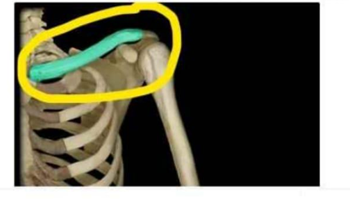
Identify the bone that is highlighted and circled in the image below.
Scapula
Clavicle
Sternum
Humerus
The Correct Answer is B
The marked structure is the clavicle, also known as the collarbone, which is a long, S-shaped bone located at the anterior base of the neck. It connects the sternum medially to the scapula laterally, forming part of the shoulder girdle. The clavicle serves as a structural strut that stabilizes the upper limb and allows free movement away from the trunk while protecting neurovascular structures beneath it.
A. Scapula: The scapula is a flat, triangular bone located on the posterior aspect of the thoracic cage, commonly referred to as the shoulder blade. It provides attachment for multiple muscles that control shoulder and arm movement, including the rotator cuff muscles. Unlike the clavicle, it is positioned on the back of the body and does not form a direct bony link to the sternum.
B. Clavicle: The clavicle is an S-shaped long bone that acts as a strut between the sternum and scapula. It stabilizes shoulder positioning and allows the upper limb to maintain a wide range of motion away from the trunk. It is the only long bone that lies horizontally in the body. Since the marked structure is anterior and connecting the shoulder girdle to the axial skeleton, the clavicle is correct.
C. Sternum: The sternum is a flat midline bone located in the anterior thorax, consisting of the manubrium, body, and xiphoid process. It serves as an attachment site for the ribs and protects vital thoracic organs such as the heart and great vessels. Unlike the clavicle, it is centrally located and does not extend laterally toward the shoulder.
D. Humerus: The humerus is the long bone of the upper arm extending from the shoulder to the elbow. It forms the major structural component of the arm and articulates with the scapula at the shoulder joint. However, it does not form the shoulder girdle connection to the sternum, making it distinct from the clavicle.
Nursing Test Bank
Naxlex Comprehensive Predictor Exams
Related Questions
Correct Answer is A
Explanation
Skeletal muscle movements are produced through coordinated interactions between different muscle groups. These muscles work in pairs or groups to create smooth, controlled motion at joints. Each muscle in a functional group has a specific role depending on whether it produces, assists, or opposes a movement. Understanding these roles is essential for interpreting biomechanics and musculoskeletal physiology.
A. Antagonist: The antagonist is the muscle that opposes or reverses the action of the agonist during movement. When one muscle contracts to produce movement, the antagonist typically relaxes to allow smooth motion, and may contract to control or decelerate the movement. For example, during elbow flexion, the triceps brachii acts as the antagonist to the biceps brachii. This opposing function helps maintain joint stability and coordinated movement.
B. Agonist: The agonist is the muscle primarily responsible for generating a specific movement. It is the main active muscle during a particular action, such as the biceps brachii during elbow flexion. The agonist contracts to produce the desired motion at a joint. Since it produces rather than opposes movement, it is not the correct answer.
C. Prime mover: The prime mover is another term for the agonist muscle, referring to the main muscle responsible for a specific movement. It generates the majority of the force required for the action. For example, the quadriceps act as the prime mover during knee extension. Because it is synonymous with agonist and not an opposing muscle, it is incorrect.
D. Synergist: A synergist is a muscle that assists the agonist in producing a movement by adding extra force or stabilizing joints. It may also prevent unwanted movements that could interfere with the primary action. For example, forearm muscles may act as synergists during hand movements. Synergists assist rather than oppose movement.
Correct Answer is D
Explanation
The wall of the eyeball is organized into three concentric layers: the outer fibrous layer, the middle vascular (uveal) layer, and the inner neural layer. The middle layer, also called the uvea, is responsible for blood supply, nourishment, and regulation of light entering the eye. It includes structures that control pupil size, lens shape, and retinal perfusion. Understanding these layers is essential for identifying ocular anatomy and related pathologies.
A. Iris: The iris is a pigmented muscular structure located in the anterior portion of the uveal tract. It contains circular (sphincter pupillae) and radial (dilator pupillae) smooth muscle fibers that regulate pupil size. This adjustment controls the amount of light entering the eye based on environmental brightness. Because it is part of the vascular middle layer, the iris is correctly included in the uvea.
B. Choroid: The choroid is a highly vascularized connective tissue layer situated between the sclera and retina. It provides oxygen and nutrient supply to the outer layers of the retina, especially the photoreceptors, which are highly metabolically active. It also absorbs excess light to prevent internal reflection within the eye. As a major component of the uveal tract, it is part of the middle eye layer.
C. Ciliary body: The ciliary body is an anterior extension of the choroid that includes the ciliary muscle and ciliary processes. It is responsible for aqueous humor production and lens accommodation by altering zonular fiber tension. This allows the lens to change shape for near and far vision focusing. Because of its vascular nature and functional integration with the iris and choroid, it is part of the middle (uveal) layer.
D. Retina: The retina is the innermost neural layer of the eye and is derived from neuroectoderm. It contains photoreceptor cells (rods and cones) that convert light energy into electrical signals through phototransduction. These signals are transmitted via bipolar and ganglion cells to the optic nerve for visual processing in the brain. Since it belongs to the inner sensory layer rather than the vascular uveal layer, it is not part of the middle eye layer.
Whether you are a student looking to ace your exams or a practicing nurse seeking to enhance your expertise , our nursing education contents will empower you with the confidence and competence to make a difference in the lives of patients and become a respected leader in the healthcare field.
Visit Naxlex, invest in your future and unlock endless possibilities with our unparalleled nursing education contents today