In this image of an ear model, name the ear structure highlighted in green and indicated by the arrow.
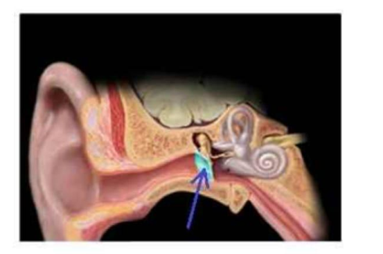
Cochlea
Tympanic membrane
Auditory ossicles
Semicircular canals
The Correct Answer is B
The marked structure is the tympanic membrane (eardrum), a thin, semi-transparent, oval-shaped membrane that separates the external auditory canal from the middle ear cavity. It plays a critical role in the conduction of sound by vibrating in response to sound waves entering the external ear. These vibrations are transmitted to the auditory ossicles (malleus, incus, and stapes), initiating mechanical amplification of sound before it reaches the inner ear. The tympanic membrane also acts as a protective barrier, preventing debris and pathogens from entering the middle ear.
A. Cochlea: The cochlea is a spiral, snail-shaped structure located in the inner ear within the petrous portion of the temporal bone. It contains the organ of Corti, which converts mechanical sound vibrations into electrical nerve impulses for hearing. Unlike the tympanic membrane, it is deeply situated and involved in sensory transduction rather than initial sound reception.
B. Tympanic membrane: The tympanic membrane is a thin, fibrous membrane that marks the boundary between the external and middle ear. It vibrates when struck by sound waves and transfers these vibrations to the malleus of the auditory ossicles. It is essential for converting airborne sound energy into mechanical energy. Since the highlighted structure is a thin circular partition at the end of the external auditory canal, it corresponds to the tympanic membrane.
C. Auditory ossicles: The auditory ossicles are three small bones (malleus, incus, and stapes) located in the middle ear cavity. Their function is to amplify and transmit sound vibrations from the tympanic membrane to the oval window of the cochlea. Unlike the tympanic membrane, they are solid bones rather than a membranous structure and are not visible as a single circular partition.
D. Semicircular canals: The semicircular canals are three looped structures of the inner ear responsible for detecting rotational movements of the head and maintaining balance. They are filled with endolymph and contain sensory receptors in the ampullae. Unlike the tympanic membrane, they are not involved in hearing but in equilibrium, and are located deeper within the temporal bone.
Nursing Test Bank
Naxlex Comprehensive Predictor Exams
Related Questions
Correct Answer is A
Explanation
The marked structure is located within smooth muscle tissue, which is composed of spindle-shaped, non-striated muscle cells found in the walls of hollow organs such as blood vessels, intestines, and the urinary bladder. These cells are responsible for involuntary movements regulated by the autonomic nervous system. In histological sections, smooth muscle is identified by elongated, spindle-shaped cells with centrally placed, cigar-shaped nuclei and absence of striations. The arrow in this image is pointing to the elongated central nucleus of a smooth muscle cell.
A. Smooth muscle cell: The image shows elongated, spindle-shaped cells arranged in parallel bundles with centrally located, elongated nuclei. These features are characteristic of smooth muscle tissue, which lacks striations due to the non-organized arrangement of actin and myosin filaments. The nucleus is the most prominent visible structure in histological sections, appearing as a dark, cigar-shaped structure in the center of each cell. Smooth muscle is responsible for involuntary contraction in organs such as blood vessels and the gastrointestinal tract, controlling processes like peristalsis and vasoconstriction.
B. Skeletal muscle cell: Skeletal muscle fibers are long, cylindrical, and multinucleated with nuclei located peripherally rather than centrally. They also display prominent cross-striations due to organized sarcomeres. Unlike the tissue shown, skeletal muscle is voluntary and found attached to bones for movement. The absence of striations and peripheral nuclei rules out skeletal muscle in this image.
C. Cardiac muscle cell: Cardiac muscle cells are branched, striated, and contain a single centrally located nucleus. They also show intercalated discs that connect adjacent cells. While they have central nuclei similar to smooth muscle, the presence of striations and branching distinguishes them. The tissue in the image lacks these features, making cardiac muscle incorrect.
D. Connective tissue fibroblast: Fibroblasts are spindle-shaped cells found in connective tissue and may appear elongated under the microscope. However, they are typically embedded within a collagen-rich extracellular matrix rather than tightly packed parallel muscle fibers. Unlike smooth muscle cells, they do not form organized contractile bundles or show uniform alignment as seen in this image.
Correct Answer is B
Explanation
The marked structure is the cervical region, which corresponds anatomically to the neck portion of the vertebral column and surrounding soft tissues. It extends from the base of the skull to the level of the first thoracic vertebra (C1–C7). This region supports the head, allows a wide range of head and neck movements, and provides a passage for critical structures including the trachea, esophagus, carotid arteries, jugular veins, and cervical spinal cord. It also contains important muscle groups such as the sternocleidomastoid and scalene muscles that contribute to posture, respiration, and head mobility.
A. Acromial region: The acromial region refers to the lateral aspect of the shoulder over the acromion process of the scapula. It forms part of the shoulder girdle and serves as an attachment point for the deltoid muscle. Unlike the cervical region, it is located on the upper lateral shoulder rather than the neck and is primarily involved in upper limb movement.
B. Cervical region: The cervical region is the neck portion of the body consisting of the cervical vertebrae and surrounding soft tissues. It supports the head, allows flexion, extension, rotation, and lateral bending, and houses vital neurovascular and airway structures. It forms the transition between the skull and thorax, making it essential for both structural support and communication pathways. Its location corresponds directly with the highlighted neck area.
C. Vertebral region: The vertebral region refers broadly to the entire spinal column, including cervical, thoracic, lumbar, sacral, and coccygeal segments. It provides axial support and protection for the spinal cord. Unlike the cervical region, it is not limited to the neck but spans the entire back from skull to pelvis.
D. Gluteal region: The gluteal region refers to the buttock area, composed mainly of the gluteus maximus, medius, and minimus muscles. It plays a key role in hip movement and locomotion. Compared to the cervical region, it is located in the lower posterior trunk and is unrelated to neck structures.
E. Lumbar region: The lumbar region refers to the lower back area containing the lumbar vertebrae (L1–L5). It supports body weight and allows trunk flexion and extension. Unlike the cervical region, it is situated between the thoracic spine and sacrum, forming the lower posterior trunk rather than the neck.
F. Sacral region: The sacral region is located at the base of the spine and consists of the fused sacral vertebrae forming the sacrum. It contributes to pelvic stability and transfers weight from the spine to the lower limbs. Compared to the cervical region, it is positioned inferiorly within the pelvis rather than in the neck.
Whether you are a student looking to ace your exams or a practicing nurse seeking to enhance your expertise , our nursing education contents will empower you with the confidence and competence to make a difference in the lives of patients and become a respected leader in the healthcare field.
Visit Naxlex, invest in your future and unlock endless possibilities with our unparalleled nursing education contents today