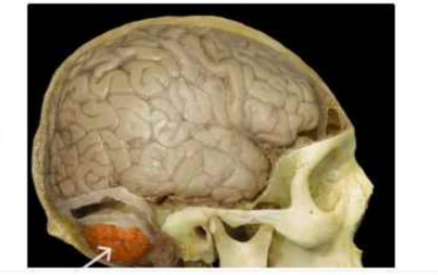
Identify the part of the brain that is highlighted and indicated by the arrow in the image below.
Cerebrum
Brainstem
Cerebellum
Medulla oblongata
The Correct Answer is C
The marked structure is the cerebellum, a major part of the hindbrain located posterior to the brainstem and inferior to the occipital lobes of the cerebrum. It consists of two hemispheres connected by the vermis and has a highly folded surface (folia) that increases its cortical area. The cerebellum is primarily responsible for coordination of voluntary movements, maintenance of posture, balance, and fine motor control. It does not initiate movement but ensures that movements are smooth, precise, and well-timed.
A. Cerebrum: The cerebrum is the largest part of the brain and consists of the cerebral hemispheres, including the frontal, parietal, temporal, and occipital lobes. It is responsible for higher cognitive functions such as reasoning, memory, language, and voluntary motor activity. Unlike the cerebellum, it is located superiorly and anteriorly in the cranial cavity and is not involved in fine motor coordination and balance regulation.
B. Brainstem: The brainstem connects the cerebrum and cerebellum to the spinal cord and consists of the midbrain, pons, and medulla oblongata. It regulates vital autonomic functions such as respiration, heart rate, and blood pressure. While it lies close to the cerebellum, it is a vertical structure inferior to the cerebrum rather than a posterior, bilobed structure like the cerebellum.
C. Cerebellum: The cerebellum is located in the posterior cranial fossa, inferior to the occipital lobes and posterior to the brainstem. It is responsible for coordinating voluntary motor activity, maintaining balance, posture, and muscle tone. It receives input from the cerebral cortex and sensory systems to fine-tune motor output. Its highly folded folia and bilateral hemispheres are characteristic features.
D. Medulla oblongata: The medulla oblongata is the lowest part of the brainstem, continuous with the spinal cord. It controls essential autonomic functions such as breathing, heart rate, and blood pressure regulation. Unlike the cerebellum, it is a narrow, tubular structure and does not have a highly folded cortical surface or function in motor coordination and balance.
Nursing Test Bank
Naxlex Comprehensive Predictor Exams
Related Questions
Correct Answer is A
Explanation
The marked structure is the scapula, a flat, triangular bone located on the posterior aspect of the thoracic cage, commonly referred to as the shoulder blade. It lies over ribs 2–7 and forms the posterior component of the shoulder girdle. The scapula plays a central role in upper limb mobility by serving as an attachment site for multiple muscles that control shoulder movement and stabilization. It articulates with the clavicle at the acromioclavicular joint and with the humerus at the glenohumeral (shoulder) joint.
A. Scapula: The scapula is a flat, triangular bone positioned on the posterior thoracic wall. It contains important anatomical landmarks such as the spine, acromion, coracoid process, and glenoid cavity, which participate in shoulder articulation and muscle attachment. It allows a wide range of shoulder movements including elevation, rotation, and abduction through coordinated muscular action. Since the marked structure lies on the posterior upper back forming the shoulder blade, it corresponds to the scapula.
B. Clavicle: The clavicle is a long, S-shaped bone located anteriorly at the base of the neck. It connects the sternum to the scapula, acting as a strut that stabilizes the shoulder girdle. Its main function is to maintain shoulder position and allow upper limb mobility away from the trunk. Unlike the scapula, it is a horizontal anterior bone rather than a flat posterior structure.
C. Humerus: The humerus is the long bone of the upper arm extending from the shoulder to the elbow joint. It serves as the main structural bone for arm movement and muscle attachment. It articulates with the scapula at the glenoid cavity to form the shoulder joint. However, it is located in the arm rather than forming the posterior shoulder blade itself.
D. Ribs: The ribs are curved, flat bones forming the thoracic cage that protects the heart and lungs. They articulate posteriorly with the thoracic vertebrae and anteriorly with the sternum (via costal cartilage). Their primary function is protection and respiratory movement. Unlike the scapula, they are part of the thoracic cage rather than the shoulder girdle.
Correct Answer is A
Explanation
Skeletal muscle contraction is based on the sliding filament theory, where thin (actin) and thick (myosin) filaments interact to produce force and movement. These interactions occur in a highly organized structural unit within myofibrils. The arrangement of sarcomeres in series allows coordinated shortening of muscle fibers. Understanding the functional unit of contraction is essential for explaining how muscles generate tension at the microscopic level.
A. Sarcomere: The sarcomere is the correct answer because it is the smallest functional (contractile) unit of skeletal muscle. It is defined as the segment between two Z-discs and contains organized actin and myosin filaments. During contraction, myosin heads bind to actin and pull the thin filaments inward, shortening the sarcomere. This coordinated shortening of many sarcomeres produces overall muscle contraction.
B. Myosin cross-bridge: a cross-bridge is a molecular interaction, not a complete functional unit. It refers specifically to the temporary attachment between a myosin head and an actin binding site. While cross-bridge cycling generates force, it occurs within the sarcomere and depends on its structural organization. It is a mechanism within the functional unit rather than the unit itself.
C. Muscle fiber: a muscle fiber is a single multinucleated muscle cell containing many myofibrils. Although it is the cellular level at which contraction occurs, it is not the smallest functional unit. Each muscle fiber contains thousands of sarcomeres arranged in series and parallel. Contraction occurs within the fiber, but the sarcomere is the true functional unit.
D. Myofibril: a myofibril is a long cylindrical structure within a muscle fiber composed of repeating sarcomeres. It serves as the structural framework for contraction but is not itself the basic contractile unit. Myofibrils transmit force generated by sarcomeres along the length of the muscle cell. It is an organizational structure rather than the functional unit of contraction.
Whether you are a student looking to ace your exams or a practicing nurse seeking to enhance your expertise , our nursing education contents will empower you with the confidence and competence to make a difference in the lives of patients and become a respected leader in the healthcare field.
Visit Naxlex, invest in your future and unlock endless possibilities with our unparalleled nursing education contents today