Identify the marked structure
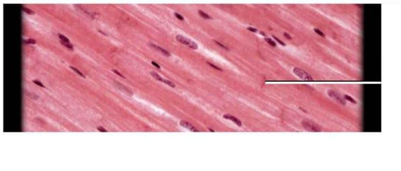
Smooth muscle
Cardiac muscle
Skeletal muscle
Connective tissue
The Correct Answer is A
The marked structure is located within smooth muscle tissue, which is composed of spindle-shaped, non-striated muscle cells found in the walls of hollow organs such as blood vessels, intestines, and the urinary bladder. These cells are responsible for involuntary movements regulated by the autonomic nervous system. In histological sections, smooth muscle is identified by elongated, spindle-shaped cells with centrally placed, cigar-shaped nuclei and absence of striations. The arrow in this image is pointing to the elongated central nucleus of a smooth muscle cell.
A. Smooth muscle cell: The image shows elongated, spindle-shaped cells arranged in parallel bundles with centrally located, elongated nuclei. These features are characteristic of smooth muscle tissue, which lacks striations due to the non-organized arrangement of actin and myosin filaments. The nucleus is the most prominent visible structure in histological sections, appearing as a dark, cigar-shaped structure in the center of each cell. Smooth muscle is responsible for involuntary contraction in organs such as blood vessels and the gastrointestinal tract, controlling processes like peristalsis and vasoconstriction.
B. Skeletal muscle cell: Skeletal muscle fibers are long, cylindrical, and multinucleated with nuclei located peripherally rather than centrally. They also display prominent cross-striations due to organized sarcomeres. Unlike the tissue shown, skeletal muscle is voluntary and found attached to bones for movement. The absence of striations and peripheral nuclei rules out skeletal muscle in this image.
C. Cardiac muscle cell: Cardiac muscle cells are branched, striated, and contain a single centrally located nucleus. They also show intercalated discs that connect adjacent cells. While they have central nuclei similar to smooth muscle, the presence of striations and branching distinguishes them. The tissue in the image lacks these features, making cardiac muscle incorrect.
D. Connective tissue fibroblast: Fibroblasts are spindle-shaped cells found in connective tissue and may appear elongated under the microscope. However, they are typically embedded within a collagen-rich extracellular matrix rather than tightly packed parallel muscle fibers. Unlike smooth muscle cells, they do not form organized contractile bundles or show uniform alignment as seen in this image.
Nursing Test Bank
Naxlex Comprehensive Predictor Exams
Related Questions
Correct Answer is C
Explanation
The marked structure is the cerebellum, a major part of the hindbrain located posterior to the brainstem and inferior to the occipital lobes of the cerebrum. It consists of two hemispheres connected by the vermis and has a highly folded surface (folia) that increases its cortical area. The cerebellum is primarily responsible for coordination of voluntary movements, maintenance of posture, balance, and fine motor control. It does not initiate movement but ensures that movements are smooth, precise, and well-timed.
A. Cerebrum: The cerebrum is the largest part of the brain and consists of the cerebral hemispheres, including the frontal, parietal, temporal, and occipital lobes. It is responsible for higher cognitive functions such as reasoning, memory, language, and voluntary motor activity. Unlike the cerebellum, it is located superiorly and anteriorly in the cranial cavity and is not involved in fine motor coordination and balance regulation.
B. Brainstem: The brainstem connects the cerebrum and cerebellum to the spinal cord and consists of the midbrain, pons, and medulla oblongata. It regulates vital autonomic functions such as respiration, heart rate, and blood pressure. While it lies close to the cerebellum, it is a vertical structure inferior to the cerebrum rather than a posterior, bilobed structure like the cerebellum.
C. Cerebellum: The cerebellum is located in the posterior cranial fossa, inferior to the occipital lobes and posterior to the brainstem. It is responsible for coordinating voluntary motor activity, maintaining balance, posture, and muscle tone. It receives input from the cerebral cortex and sensory systems to fine-tune motor output. Its highly folded folia and bilateral hemispheres are characteristic features.
D. Medulla oblongata: The medulla oblongata is the lowest part of the brainstem, continuous with the spinal cord. It controls essential autonomic functions such as breathing, heart rate, and blood pressure regulation. Unlike the cerebellum, it is a narrow, tubular structure and does not have a highly folded cortical surface or function in motor coordination and balance.
Correct Answer is D
Explanation
Muscle contraction at the cellular level is explained by the sliding filament theory, which involves the interaction between thick and thin filaments within the sarcomere. Thin filaments are essential structural components of skeletal and cardiac muscle fibers and play a key role in generating force during contraction. They are anchored to the Z-line and interact with thick filaments (myosin) to produce shortening of the sarcomere. The thin filament complex is composed of multiple proteins that regulate and facilitate contraction.
A. Myosin: Myosin is the primary protein of thick filaments, not thin filaments. It functions as a motor protein with ATPase activity, allowing it to bind to actin and generate the power stroke that produces muscle contraction. The myosin heads form cross-bridges with actin during contraction. Since it belongs to thick filaments, it is not the main component of thin filaments.
B. Troponin:Troponin is a regulatory protein complex located on thin filaments. It consists of three subunits: troponin C (binds calcium), troponin I (inhibits actin-myosin interaction), and troponin T (binds tropomyosin). Its role is to regulate the exposure of myosin-binding sites on actin in response to calcium levels. Although essential for contraction regulation, it is not the main structural component of thin filaments.
C. Acetylcholine: Acetylcholine is a neurotransmitter released at the neuromuscular junction, not a structural component of muscle filaments. It binds to receptors on the muscle fiber membrane (sarcolemma) to initiate depolarization and trigger muscle contraction. Its function is purely chemical signaling between nerve and muscle. Therefore, it is not part of the thin filament structure.
D. Actin: Actin is the primary structural protein of thin filaments in muscle cells. It forms a helical chain of globular actin (G-actin) that polymerizes into filamentous actin (F-actin), providing binding sites for myosin heads during contraction. Actin works together with regulatory proteins such as tropomyosin and troponin to control contraction. Because it forms the core structural backbone of thin filaments, it is the correct answer.
Whether you are a student looking to ace your exams or a practicing nurse seeking to enhance your expertise , our nursing education contents will empower you with the confidence and competence to make a difference in the lives of patients and become a respected leader in the healthcare field.
Visit Naxlex, invest in your future and unlock endless possibilities with our unparalleled nursing education contents today