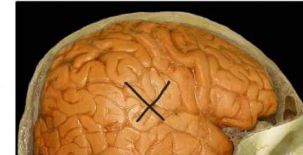
Identify the part of the brain that is highlighted and indicated by an X in the image below.
Occipital lobe
Frontal lobe
Parietal lobe
Temporal lobe
The Correct Answer is D
The marked structure is the temporal lobe, one of the four major lobes of the cerebral cortex located on the lateral and inferior aspect of each cerebral hemisphere, beneath the lateral (Sylvian) fissure. It is structurally composed of multiple gyri and sulci that increase cortical surface area for higher processing capacity. The temporal lobe plays a crucial role in auditory processing, language comprehension (Wernicke’s area in the dominant hemisphere), memory formation via the hippocampal connections, and emotional responses through limbic system integration.
A. Occipital lobe: The occipital lobe is located at the posterior aspect of the cerebral hemispheres and is primarily responsible for visual processing. It contains the primary visual cortex (V1), which interprets input from the retina via the optic pathways. Unlike the temporal lobe, it does not process auditory information or language comprehension. Its position at the back of the brain also distinguishes it from the lateral location of the temporal lobe.
B. Frontal lobe: The frontal lobe is located in the anterior portion of the cerebral hemisphere and is responsible for executive functions such as reasoning, planning, voluntary motor control, and speech production (Broca’s area). It also regulates personality, judgment, and emotional control. Compared to the temporal lobe, it is more anterior and superior, and is not primarily involved in auditory perception or memory consolidation.
C. Parietal lobe: The parietal lobe is located superiorly on the cerebral hemisphere and is mainly responsible for somatosensory processing, including touch, temperature, pain, and proprioception. It integrates sensory input to form spatial awareness and body orientation. Unlike the temporal lobe, it is positioned superiorly and is not directly involved in auditory processing or memory systems.
D. Temporal lobe: The temporal lobe is located on the lateral aspect of the brain, inferior to the lateral sulcus. It contains the primary auditory cortex and is essential for processing sound, language comprehension, and memory encoding via hippocampal connections. It also plays a role in emotional regulation through limbic system interactions. Since the marked area is lateral and associated with auditory and language functions, it corresponds to the temporal lobe.
Nursing Test Bank
Naxlex Comprehensive Predictor Exams
Related Questions
Correct Answer is C
Explanation
The marked structure is the dendrite, a branched projection of a neuron that extends from the cell body (soma). Dendrites are specialized for receiving synaptic input from other neurons and transmitting that electrical signal toward the cell body. They increase the surface area available for synaptic connections, allowing integration of multiple incoming signals. Functionally, dendrites play a key role in determining whether a neuron reaches the threshold for action potential generation at the axon hillock.
A. Axon terminal: The axon terminal is the distal end of the axon where neurotransmitters are released into the synaptic cleft. It forms synapses with other neurons, muscles, or glands to transmit signals to the next cell. Unlike dendrites, which receive input, axon terminals are involved in signal output. They are usually found at the far end of long axonal projections rather than branching near the cell body.
B. Axon: The axon is a long, single projection that carries electrical impulses away from the neuron’s cell body toward target cells. It is often myelinated to increase conduction speed and ends in terminal branches. Unlike dendrites, it is typically singular, longer, and specialized for signal transmission rather than reception.
C. Dendrite: Dendrites are multiple, short, highly branched extensions of the neuron that receive incoming synaptic signals. They conduct graded potentials toward the soma, where integration occurs to determine neuronal firing. Their extensive branching increases receptive surface area, making them essential for neural communication. Their structure and function as primary input receivers make them the correct answer.
D. Cell body: The cell body (soma) contains the nucleus and most organelles required for neuronal metabolism and protein synthesis. It integrates incoming signals from dendrites and maintains cell function. Unlike dendrites, it is not a branching structure but a central region of the neuron. It serves as the metabolic center rather than the primary input surface.
Correct Answer is D
Explanation
The wall of the eyeball is organized into three concentric layers: the outer fibrous layer, the middle vascular (uveal) layer, and the inner neural layer. The middle layer, also called the uvea, is responsible for blood supply, nourishment, and regulation of light entering the eye. It includes structures that control pupil size, lens shape, and retinal perfusion. Understanding these layers is essential for identifying ocular anatomy and related pathologies.
A. Iris: The iris is a pigmented muscular structure located in the anterior portion of the uveal tract. It contains circular (sphincter pupillae) and radial (dilator pupillae) smooth muscle fibers that regulate pupil size. This adjustment controls the amount of light entering the eye based on environmental brightness. Because it is part of the vascular middle layer, the iris is correctly included in the uvea.
B. Choroid: The choroid is a highly vascularized connective tissue layer situated between the sclera and retina. It provides oxygen and nutrient supply to the outer layers of the retina, especially the photoreceptors, which are highly metabolically active. It also absorbs excess light to prevent internal reflection within the eye. As a major component of the uveal tract, it is part of the middle eye layer.
C. Ciliary body: The ciliary body is an anterior extension of the choroid that includes the ciliary muscle and ciliary processes. It is responsible for aqueous humor production and lens accommodation by altering zonular fiber tension. This allows the lens to change shape for near and far vision focusing. Because of its vascular nature and functional integration with the iris and choroid, it is part of the middle (uveal) layer.
D. Retina: The retina is the innermost neural layer of the eye and is derived from neuroectoderm. It contains photoreceptor cells (rods and cones) that convert light energy into electrical signals through phototransduction. These signals are transmitted via bipolar and ganglion cells to the optic nerve for visual processing in the brain. Since it belongs to the inner sensory layer rather than the vascular uveal layer, it is not part of the middle eye layer.
Whether you are a student looking to ace your exams or a practicing nurse seeking to enhance your expertise , our nursing education contents will empower you with the confidence and competence to make a difference in the lives of patients and become a respected leader in the healthcare field.
Visit Naxlex, invest in your future and unlock endless possibilities with our unparalleled nursing education contents today