Identify the muscle of the eye indicated by the arrow in the image below.
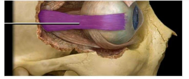
Lateral rectus
Medial rectus
Superior rectus
Inferior oblique
The Correct Answer is A
The marked structure is the lateral rectus muscle, one of the six extraocular muscles responsible for eye movement. It originates from the annular tendon (common tendinous ring) at the apex of the orbit and inserts on the lateral aspect of the sclera. Its primary physiological function is abduction of the eyeball, meaning it moves the eye outward away from the midline. It is innervated by the abducens nerve (CN VI), which is clinically important because lesions result in inability to move the eye laterally and horizontal diplopia.
A. Lateral rectus: The lateral rectus is a straight extraocular muscle located on the lateral side of the orbit. It functions exclusively to abduct the eye, pulling the eyeball outward toward the temporal side. It is innervated by cranial nerve VI (abducens nerve), making it vulnerable in increased intracranial pressure. Its lateral position and abduction function match the indicated structure.
B. Medial rectus: The medial rectus is located on the medial side of the orbit and is responsible for adduction of the eye, moving it toward the nasal side. It is innervated by the oculomotor nerve (CN III). Unlike the lateral rectus, it pulls the eye inward rather than outward. Its position opposite the lateral rectus helps coordinate horizontal eye movements but does not match the lateral positioning in the image.
C. Superior rectus: The superior rectus is located on the superior aspect of the orbit and elevates the eyeball. It also contributes to adduction and intorsion depending on eye position. It is innervated by CN III. Unlike the lateral rectus, it is not responsible for horizontal abduction and is positioned above the globe rather than on the lateral side.
D. Inferior oblique: The inferior oblique is a curved muscle originating from the anterior medial orbital floor and inserting on the posterolateral sclera. It elevates and externally rotates the eye, especially when the eye is adducted. It is unique in its oblique course and is not a straight lateral muscle. Compared to the lateral rectus, it is deeper and functions mainly in vertical and rotational movements rather than abduction.
Nursing Test Bank
Naxlex Comprehensive Predictor Exams
Related Questions
Correct Answer is D
Explanation
Sensory receptors are specialized structures that detect specific environmental or internal stimuli and convert them into electrical signals for the central nervous system. They are classified based on the type of stimulus they respond to, such as chemical, thermal, light, or mechanical changes. Mechanoreceptors are particularly important in detecting physical deformation such as stretch, pressure, vibration, and tension. Stretch receptors and baroreceptors fall within this category because they respond to mechanical distortion of tissues and blood vessels.
A. Chemoreceptors: Chemoreceptors detect chemical changes in the internal or external environment, such as oxygen, carbon dioxide, pH levels, and specific dissolved substances. They are found in structures like the carotid bodies, aortic bodies, and taste buds. Their function is essential in regulating respiration and maintaining acid-base balance. Stretch receptors and baroreceptors do not respond to chemical changes.
B. Thermoreceptors: Thermoreceptors are sensory receptors that detect changes in temperature, both heat and cold. They are primarily located in the skin and hypothalamus and help regulate body temperature through autonomic responses such as sweating or shivering. Their function is related to thermal homeostasis rather than pressure or stretch detection.
C. Photoreceptors: Photoreceptors are specialized sensory cells located in the retina that respond to light stimuli. They include rods, which detect low light intensity, and cones, which are responsible for color vision and visual acuity. Their function is essential for vision and image formation. Since they respond to electromagnetic light waves rather than mechanical deformation, they are not related to stretch receptors or baroreceptors.
D. Mechanoreceptors: Mechanoreceptors are sensory receptors that respond to mechanical stimuli such as pressure, stretch, vibration, and tension. Stretch receptors located in muscles and lungs detect changes in length, while baroreceptors in the carotid sinus and aortic arch detect changes in blood vessel wall pressure. These receptors play a key role in regulating blood pressure, proprioception, and visceral reflexes. Because they respond specifically to mechanical deformation, stretch receptors and baroreceptors are correctly classified as mechanoreceptors.
Correct Answer is C
Explanation
The marked structure is the mandible, which is the largest and strongest facial bone forming the lower jaw. It is the only movable bone of the skull, articulating with the temporal bone at the temporomandibular joint (TMJ). The mandible supports the lower teeth and plays a critical role in mastication, speech, and facial structure. It is shaped like a horseshoe and consists of the body and two rami.
A. Maxilla: The maxilla forms the upper jaw and is a fixed bone of the facial skeleton. It contributes to the hard palate, the floor of the orbit, and the upper dental arch. Unlike the mandible, it is immovable and does not form a joint for chewing motion.
B. Zygomatic bone: The zygomatic bone forms the prominence of the cheek and part of the lateral wall of the orbit. It contributes to facial contour and protection of the eye. Compared to the mandible, it is a non-movable facial bone and does not participate in jaw movement or mastication.
C. Mandible: The mandible is the lower jawbone and the only movable bone of the skull. It articulates with the temporal bone at the TMJ, allowing chewing, speaking, and mouth opening. It supports the lower teeth and provides attachment for muscles of mastication such as the masseter and temporalis. Since the marked structure is the movable lower jaw, it corresponds to the mandible.
D. Temporal bone: The temporal bone forms part of the lateral skull and houses structures of the ear. It contributes to the cranial base and forms the socket for the mandibular articulation (TMJ). However, it is not the jawbone itself but rather the bone that articulates with the mandible.
Whether you are a student looking to ace your exams or a practicing nurse seeking to enhance your expertise , our nursing education contents will empower you with the confidence and competence to make a difference in the lives of patients and become a respected leader in the healthcare field.
Visit Naxlex, invest in your future and unlock endless possibilities with our unparalleled nursing education contents today