Identify the layer of the skin indicated by the line.
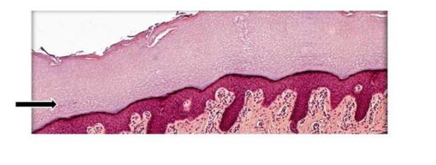
Dermis
Hypodermis
Epidermis
Reticular layer
The Correct Answer is C
The marked structure is the epidermis, the most superficial layer of the skin forming the outer protective barrier of the body. It is composed primarily of stratified squamous keratinized epithelium and is avascular, relying on diffusion from the underlying dermis for nutrient supply. The epidermis is responsible for preventing water loss, blocking pathogen entry, and protecting against mechanical, chemical, and UV damage. It also contains specialized cells such as keratinocytes, melanocytes, Langerhans cells, and Merkel cells that contribute to barrier function, pigmentation, immune defense, and sensory perception.
A. Dermis: The dermis is the thick, connective tissue layer beneath the epidermis that provides structural support and elasticity to the skin. It contains collagen and elastin fibers, blood vessels, nerve endings, hair follicles, and sweat glands. Unlike the epidermis, it is vascular and plays a major role in thermoregulation and nutrient supply to the epidermis.
B. Hypodermis: The hypodermis (subcutaneous layer) lies beneath the dermis and is composed mainly of adipose and loose connective tissue. It functions in energy storage, insulation, and cushioning of underlying structures. Compared to the epidermis, it is much deeper and does not form the external protective barrier of the skin.
C. Epidermis: The epidermis is the outermost skin layer composed of stratified squamous keratinized epithelium. It provides the primary barrier against environmental injury, dehydration, and microbial invasion. It undergoes continuous regeneration through basal cell division and keratinization as cells move toward the surface. Its superficial location and protective role make it the correct identification.
D. Reticular layer: The reticular layer is the deeper portion of the dermis composed of dense irregular connective tissue. It provides strength and elasticity to the skin due to its thick collagen fiber network. Unlike the epidermis, it is not the outermost layer and does not directly interact with the external environment.
Nursing Test Bank
Naxlex Comprehensive Predictor Exams
Related Questions
Correct Answer is C
Explanation
The facial skeleton is composed of several paired and unpaired bones that form the architecture of the orbits, nasal cavity, and oral cavity. The bone marked with an "X" in represents a central structural element of the midface, serving as the foundation for the upper teeth and contributing to the floor of the nasal cavity. Understanding the complex articulations of these facial bones is critical for clinicians evaluating facial structure, dental alignment, and potential fractures of the midface region.
A. The nasal bones are a pair of small, rectangular bones that form the bridge of the nose. They are located superior to the area marked with an "X" . While they are part of the nasal region, the marked area is situated lower, on the alveolar process of the upper jaw, which is distinct from the nasal bridge.
B. The vomer is an unpaired, thin, plow-shaped bone that forms the posterior and inferior part of the nasal septum. It is situated deep within the nasal cavity and is not visible from this external anterior view of the skull. The mark "X" is placed on the external surface of the facial skeleton, not deep within the midline nasal structure.
C. The maxilla is the correct identification for the structure marked with an "X". This bone forms the entire upper jaw, the majority of the hard palate, and the lower margins of the nasal aperture. The area indicated is the alveolar process of the maxilla, which contains the sockets (alveoli) for the upper teeth, confirming its role as the primary bone of the midface.
D. The mandible is the lower jaw bone and is the only mobile bone of the skull. It is located inferior to the maxilla and is separated from it by the oral cavity. Because the area marked with the "X" is firmly attached to the midface region above the upper teeth, it is anatomically separate from the mandible.
Correct Answer is C
Explanation
Acetylcholine (ACh) is a key neurotransmitter involved in both the central and peripheral nervous systems, particularly at neuromuscular junctions and autonomic synapses. After ACh is released into the synaptic cleft, its action must be rapidly terminated to allow precise control of nerve signaling and prevent continuous stimulation. This termination is achieved by enzymatic degradation. Acetylcholinesterase is the enzyme responsible for this process, ensuring proper synaptic function and muscle relaxation.
A. To synthesize acetylcholine: acetylcholine is synthesized by the enzyme choline acetyltransferase (ChAT), not acetylcholinesterase. ChAT combines choline and acetyl-CoA within the presynaptic neuron to form acetylcholine. Acetylcholinesterase functions after release, not during synthesis.
B. To transport acetylcholine across the synaptic cleft: acetylcholine is not actively transported across the synaptic cleft. Instead, it is released by exocytosis from presynaptic vesicles and diffuses passively across the synaptic gap to bind receptors on the postsynaptic membrane. No transport protein carries it across the cleft.
C. To break down acetylcholine into acetate and choline: acetylcholinesterase rapidly hydrolyzes acetylcholine in the synaptic cleft into acetate and choline. This enzymatic breakdown terminates the signal at cholinergic synapses, preventing continuous stimulation of the postsynaptic receptor. The choline produced is then recycled back into the presynaptic neuron for resynthesis of acetylcholine. This mechanism ensures precise and rapid control of neural transmission.
D. To increase the release of dopamine at the synapse: acetylcholinesterase is specific to acetylcholine and does not influence dopamine release. Dopamine release is regulated by different enzymes and transport mechanisms within dopaminergic neurons. Acetylcholinesterase has no role in modulating dopamine levels or synaptic release.
Whether you are a student looking to ace your exams or a practicing nurse seeking to enhance your expertise , our nursing education contents will empower you with the confidence and competence to make a difference in the lives of patients and become a respected leader in the healthcare field.
Visit Naxlex, invest in your future and unlock endless possibilities with our unparalleled nursing education contents today