Identify the shape of the cell indicated by the arrow.
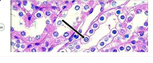
Squamous
Cuboidal
Columnar
Stratified
The Correct Answer is B
The cell structure represents cuboidal epithelial cells, which are characterized by their cube-like shape with approximately equal height and width and a centrally located, round nucleus. Cuboidal epithelium is commonly found in glandular tissue and kidney tubules, where it plays an important role in secretion and absorption. Its structural design provides a balance between surface area and cellular volume, allowing efficient metabolic and transport activities.
A. Squamous: Squamous cells are thin, flat, and scale-like in shape with flattened nuclei. They are specialized for diffusion and filtration due to their minimal thickness, as seen in structures like alveoli and capillary endothelium. Unlike cuboidal cells, they are not cube-shaped and are less involved in active secretion or absorption.
B. Cuboidal: Cuboidal cells are roughly square-shaped epithelial cells with centrally placed nuclei. They are well adapted for secretion and absorption due to a higher cytoplasmic volume compared to squamous cells. They commonly line kidney tubules, small ducts of glands, and thyroid follicles. Their equal height and width make them the correct identification.
C. Columnar: Columnar cells are tall, rectangular epithelial cells with nuclei typically located toward the basal region. They are specialized for absorption and secretion and often contain microvilli or cilia depending on location. Compared to cuboidal cells, they are taller than they are wide and are commonly found in the gastrointestinal tract rather than kidney tubules.
D. Stratified: Stratified epithelium refers to multiple layers of epithelial cells rather than a single cell shape. It is classified based on arrangement, not individual cell morphology. Its main function is protection against abrasion in areas like the skin and oral cavity. Unlike cuboidal cells, it does not describe a single cell shape but a tissue organization pattern.
Nursing Test Bank
Naxlex Comprehensive Predictor Exams
Related Questions
Correct Answer is D
Explanation
The marked structure is the temporal lobe, one of the four major lobes of the cerebral cortex located on the lateral and inferior aspect of each cerebral hemisphere, beneath the lateral (Sylvian) fissure. It is structurally composed of multiple gyri and sulci that increase cortical surface area for higher processing capacity. The temporal lobe plays a crucial role in auditory processing, language comprehension (Wernicke’s area in the dominant hemisphere), memory formation via the hippocampal connections, and emotional responses through limbic system integration.
A. Occipital lobe: The occipital lobe is located at the posterior aspect of the cerebral hemispheres and is primarily responsible for visual processing. It contains the primary visual cortex (V1), which interprets input from the retina via the optic pathways. Unlike the temporal lobe, it does not process auditory information or language comprehension. Its position at the back of the brain also distinguishes it from the lateral location of the temporal lobe.
B. Frontal lobe: The frontal lobe is located in the anterior portion of the cerebral hemisphere and is responsible for executive functions such as reasoning, planning, voluntary motor control, and speech production (Broca’s area). It also regulates personality, judgment, and emotional control. Compared to the temporal lobe, it is more anterior and superior, and is not primarily involved in auditory perception or memory consolidation.
C. Parietal lobe: The parietal lobe is located superiorly on the cerebral hemisphere and is mainly responsible for somatosensory processing, including touch, temperature, pain, and proprioception. It integrates sensory input to form spatial awareness and body orientation. Unlike the temporal lobe, it is positioned superiorly and is not directly involved in auditory processing or memory systems.
D. Temporal lobe: The temporal lobe is located on the lateral aspect of the brain, inferior to the lateral sulcus. It contains the primary auditory cortex and is essential for processing sound, language comprehension, and memory encoding via hippocampal connections. It also plays a role in emotional regulation through limbic system interactions. Since the marked area is lateral and associated with auditory and language functions, it corresponds to the temporal lobe.
Correct Answer is C
Explanation
Broca’s area is a specialized region of the dominant frontal lobe (usually the left hemisphere) located in the inferior frontal gyrus. It plays a critical role in the motor production of speech and language formulation. This area coordinates the complex motor planning required to articulate words, allowing fluent verbal expression. Damage to this region disrupts speech output while often preserving comprehension, resulting in a characteristic language deficit known as expressive aphasia.
A. Memory loss: memory processing is primarily associated with the hippocampus and medial temporal lobe structures. These areas are responsible for forming and retrieving long-term memories. Broca’s area does not play a direct role in memory storage or recall. Therefore, damage to Broca’s area would not primarily result in memory impairment.
B. Impaired motor control of the left side of the body: motor control of the body is governed by the primary motor cortex in the frontal lobe, particularly the precentral gyrus. Additionally, motor pathways decussate in the brainstem, meaning the right hemisphere controls the left side of the body. While nearby motor regions may be involved in movement, Broca’s area is specifically dedicated to speech production, not general motor control.
C. Inability to produce speech (expressive aphasia): Broca’s area is responsible for the motor planning and production of speech. Damage to this region results in expressive aphasia, where individuals understand language but struggle to form coherent spoken or written speech. Speech becomes slow, effortful, and fragmented, although comprehension remains relatively intact. This demonstrates the specialized role of Broca’s area in language expression.
D. Loss of visual processing: visual processing occurs in the occipital lobe, specifically the primary visual cortex. This region interprets visual stimuli received from the retina via the optic pathways. Broca’s area has no involvement in visual perception or interpretation. Therefore, damage to this area would not affect vision.
Whether you are a student looking to ace your exams or a practicing nurse seeking to enhance your expertise , our nursing education contents will empower you with the confidence and competence to make a difference in the lives of patients and become a respected leader in the healthcare field.
Visit Naxlex, invest in your future and unlock endless possibilities with our unparalleled nursing education contents today