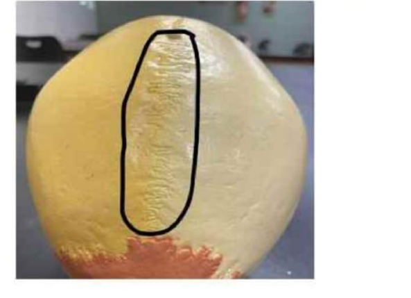
Identify the suture circled in the image below.
Coronal suture
Lambdoid suture
Squamous suture
Sagittal suture
The Correct Answer is D
The marked structure is the sagittal suture, a fibrous immovable joint (synarthrosis) located between the two parietal bones along the midline of the superior skull. It runs anteroposteriorly from the coronal suture anteriorly to the lambdoid suture posteriorly. The sagittal suture plays a key role in skull growth during childhood by allowing expansion of the cranial vault to accommodate brain development. It later ossifies progressively in adulthood, becoming more rigid.
A. Coronal suture: The coronal suture is located between the frontal bone and the paired parietal bones. It runs transversely across the skull from side to side, separating the anterior cranial vault from the superior region. It allows growth of the skull in the anterior-posterior direction during early development. Unlike the sagittal suture, it is not located along the midline but rather forms a horizontal junction.
B. Lambdoid suture: The lambdoid suture is found at the posterior aspect of the skull, where the parietal bones meet the occipital bone. It has a characteristic inverted “V” or lambda shape. It plays a role in posterior cranial expansion during development. Compared to the sagittal suture, it is posterior and not located along the midline of the skull.
C. Squamous suture: The squamous suture is located laterally between the temporal bone and the parietal bone. It is curved and relatively flat, forming part of the lateral skull wall. It allows articulation between these bones and contributes to cranial stability. It is not midline like the sagittal suture and is instead positioned on the sides of the skull.
D. Sagittal suture: The sagittal suture is a fibrous joint located along the midline between the two parietal bones. It extends from the coronal suture anteriorly to the lambdoid suture posteriorly. It is a key growth site during infancy and childhood, allowing expansion of the skull to accommodate brain growth. Since the circled structure lies centrally between the parietal bones, it corresponds to the sagittal suture.
Nursing Test Bank
Naxlex Comprehensive Predictor Exams
Related Questions
Correct Answer is B
Explanation
The marked structure is the cervical region, which corresponds anatomically to the neck portion of the vertebral column and surrounding soft tissues. It extends from the base of the skull to the level of the first thoracic vertebra (C1–C7). This region supports the head, allows a wide range of head and neck movements, and provides a passage for critical structures including the trachea, esophagus, carotid arteries, jugular veins, and cervical spinal cord. It also contains important muscle groups such as the sternocleidomastoid and scalene muscles that contribute to posture, respiration, and head mobility.
A. Acromial region: The acromial region refers to the lateral aspect of the shoulder over the acromion process of the scapula. It forms part of the shoulder girdle and serves as an attachment point for the deltoid muscle. Unlike the cervical region, it is located on the upper lateral shoulder rather than the neck and is primarily involved in upper limb movement.
B. Cervical region: The cervical region is the neck portion of the body consisting of the cervical vertebrae and surrounding soft tissues. It supports the head, allows flexion, extension, rotation, and lateral bending, and houses vital neurovascular and airway structures. It forms the transition between the skull and thorax, making it essential for both structural support and communication pathways. Its location corresponds directly with the highlighted neck area.
C. Vertebral region: The vertebral region refers broadly to the entire spinal column, including cervical, thoracic, lumbar, sacral, and coccygeal segments. It provides axial support and protection for the spinal cord. Unlike the cervical region, it is not limited to the neck but spans the entire back from skull to pelvis.
D. Gluteal region: The gluteal region refers to the buttock area, composed mainly of the gluteus maximus, medius, and minimus muscles. It plays a key role in hip movement and locomotion. Compared to the cervical region, it is located in the lower posterior trunk and is unrelated to neck structures.
E. Lumbar region: The lumbar region refers to the lower back area containing the lumbar vertebrae (L1–L5). It supports body weight and allows trunk flexion and extension. Unlike the cervical region, it is situated between the thoracic spine and sacrum, forming the lower posterior trunk rather than the neck.
F. Sacral region: The sacral region is located at the base of the spine and consists of the fused sacral vertebrae forming the sacrum. It contributes to pelvic stability and transfers weight from the spine to the lower limbs. Compared to the cervical region, it is positioned inferiorly within the pelvis rather than in the neck.
Correct Answer is C
Explanation
During embryonic development, the central nervous system originates from three primary brain vesicles: the forebrain (prosencephalon), midbrain (mesencephalon), and hindbrain (rhombencephalon). Each of these vesicles differentiates into specific structures of the mature brain. The forebrain is the most complex and gives rise to higher-order processing centers involved in cognition, sensory integration, and autonomic regulation. It ultimately forms both the cerebrum and diencephalon, which are essential for conscious thought and homeostatic control.
A. Medulla oblongata and spinal cord: these structures develop from the hindbrain (rhombencephalon) and neural tube, not the forebrain. The medulla oblongata is part of the brainstem and is responsible for autonomic functions such as respiration and heart rate regulation. The spinal cord arises caudal to the brainstem from the neural tube. Therefore, they are not derivatives of the forebrain.
B. Cerebellum and pons: both the cerebellum and pons arise from the hindbrain, specifically the metencephalon. The cerebellum is responsible for coordination and balance, while the pons acts as a relay center between different parts of the brain. These structures are not derived from the embryonic forebrain.
C. Cerebrum and diencephalon: the embryonic forebrain (prosencephalon) differentiates into the telencephalon and diencephalon. The telencephalon develops into the cerebrum, which is responsible for higher cognitive functions, voluntary movement, and sensory perception. The diencephalon forms structures such as the thalamus and hypothalamus, which are involved in sensory relay and autonomic regulation. These structures collectively represent the mature derivatives of the forebrain.
D. Midbrain and medulla oblongata: the midbrain develops from the mesencephalon (midbrain vesicle), and the medulla oblongata develops from the hindbrain. The forebrain does not contribute to either of these structures.
Whether you are a student looking to ace your exams or a practicing nurse seeking to enhance your expertise , our nursing education contents will empower you with the confidence and competence to make a difference in the lives of patients and become a respected leader in the healthcare field.
Visit Naxlex, invest in your future and unlock endless possibilities with our unparalleled nursing education contents today