Identify the structure shown below
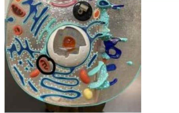
Tissue
Cell
Organ
Organ system
The Correct Answer is B
The structure shown represents a cell, which is the fundamental structural and functional unit of life. Cells are the smallest living units capable of carrying out essential life processes such as metabolism, growth, reproduction, and response to stimuli. All tissues, organs, and organ systems are ultimately composed of specialized cells working together in coordinated ways. Cells vary widely in structure depending on their function, but all share basic components such as the plasma membrane, cytoplasm, and genetic material.
A. Tissue: A tissue is a group of similar cells working together to perform a specific function, such as epithelial, connective, muscle, or nervous tissue. Tissues are more complex than individual cells and form the structural basis of organs. Unlike a single cell, tissues show organized layers or clusters of multiple cells performing collective functions. Therefore, a tissue is a higher level of biological organization than the structure shown.
B. Cell: A cell is the smallest independent unit of life and the basic building block of all living organisms. It contains specialized organelles such as the nucleus, mitochondria, and endoplasmic reticulum, which carry out metabolic and regulatory functions. Cells maintain homeostasis, produce energy, and carry genetic information necessary for replication.
C. Organ: An organ is composed of two or more different tissues working together to perform a specific function, such as the heart, lungs, or kidneys. Organs are more complex than cells and tissues and contribute to broader physiological processes in the body. Unlike a single cell, organs have macroscopic structure and coordinated systems of function.
D. Organ system: An organ system consists of multiple organs working together to carry out major bodily functions, such as the respiratory, digestive, or nervous systems. It represents a highly complex level of organization above organs and tissues. Organ systems coordinate large-scale physiological processes essential for survival. Compared to a single cell, an organ system is vastly more complex and structurally extensive.
Nursing Test Bank
Naxlex Comprehensive Predictor Exams
Related Questions
Correct Answer is C
Explanation
Light entering the eye follows a precise anatomical pathway through transparent refractive media before reaching the retina for phototransduction. These structures work together to bend (refract) and focus light onto the photoreceptor layer to produce a clear image. The cornea provides the greatest refractive power, while the lens fine-tunes focusing. The aqueous and vitreous humors maintain intraocular pressure and allow unobstructed transmission of light.
A. Vitreous humor → lens → aqueous humor → cornea: This sequence reverses the normal anterior-to-posterior direction of light entry into the eye. Light first encounters the cornea, not the vitreous humor, which is located in the posterior segment of the eye. The vitreous humor lies behind the lens and cannot be the initial medium for light transmission.
B. Cornea → lens → aqueous humor → vitreous humor: This misplaces the aqueous humor after the lens. Anatomically, aqueous humor is located between the cornea and lens, filling both the anterior and posterior chambers. Light must pass through aqueous humor before reaching the lens, not after it. This sequence disrupts the correct spatial arrangement of the eye’s refractive media.
C. Cornea → aqueous humor → lens → vitreous humor: Light first enters the cornea, which provides the majority of refractive power due to its curved structure and air–cornea interface. It then passes through the aqueous humor in the anterior chamber, followed by the lens, which adjusts focus through accommodation. Finally, light travels through the vitreous humor before reaching the retina for image formation.
D. Aqueous humor → cornea → lens → vitreous humor: This option places aqueous humor before the cornea, which is anatomically inaccurate. The cornea is the first structure encountered by incoming light and serves as the primary refractive surface. Aqueous humor lies posterior to the cornea and cannot precede it in the light pathway. This sequence misrepresents the anatomical organization of the anterior segment of the eye.
Correct Answer is B
Explanation
Skeletal muscle fibers are highly specialized cells designed for rapid and coordinated contraction. To achieve this, they require an efficient system for transmitting electrical signals from the cell surface deep into the muscle fiber. Transverse (T) tubules are invaginations of the sarcolemma that penetrate into the cell interior. They work closely with the sarcoplasmic reticulum to ensure uniform and rapid activation of muscle contraction throughout the fiber.
A. To store calcium ions needed for activating tropomyosin: calcium storage in muscle cells is primarily handled by the sarcoplasmic reticulum, not the T-tubules. The sarcoplasmic reticulum releases calcium ions in response to an action potential, allowing calcium to bind troponin and shift tropomyosin away from actin binding sites. T-tubules do not store calcium; they serve as conduits for electrical signals.
B. To transmit action potentials (impulses) into the cell interior: transverse tubules are invaginations of the sarcolemma that rapidly conduct action potentials from the cell surface into the deeper regions of the muscle fiber. This ensures that the entire muscle fiber contracts simultaneously rather than in a wave-like fashion. The electrical signal traveling through T-tubules triggers calcium release from the sarcoplasmic reticulum. This coupling is essential for coordinated and efficient muscle contraction.
C. To synthesize ATP for muscle contraction: ATP production occurs primarily in mitochondria through oxidative phosphorylation and in the cytoplasm via glycolysis. T-tubules have no role in energy metabolism or ATP synthesis. Their function is electrical signal transmission, not biochemical energy production. Therefore, this option describes a mitochondrial function rather than a T-tubule function.
D. To break down acetylcholine at the neuromuscular junction: acetylcholine breakdown is performed by the enzyme acetylcholinesterase located in the synaptic cleft of the neuromuscular junction. T-tubules are located inside the muscle fiber and are not involved in synaptic transmission or neurotransmitter degradation.
Whether you are a student looking to ace your exams or a practicing nurse seeking to enhance your expertise , our nursing education contents will empower you with the confidence and competence to make a difference in the lives of patients and become a respected leader in the healthcare field.
Visit Naxlex, invest in your future and unlock endless possibilities with our unparalleled nursing education contents today