In the diagram above #3 indicates
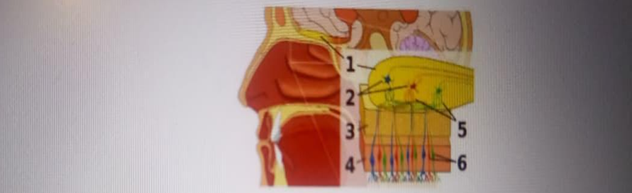
olfactory bulb
olfactory stem cells
fibers of the olfactory nerves
cribiform plate of ethmoid bone
The Correct Answer is D
A. olfactory bulb: This structure, labeled as #1 in the diagram, is the distal part of the olfactory tract where the first-order olfactory neurons synapse with second-order mitral cells. It sits directly above the bony floor of the anterior cranial fossa and serves as the initial processing center for odorant information before it is sent to the primary olfactory cortex.
B. olfactory stem cells: Also known as basal cells, are located at the base of the olfactory epithelium and serve as regenerative precursors for the specialized olfactory receptor neurons. They are essential for maintaining the olfactory system's function because the sensory neurons are frequently exposed to environmental toxins and must be replaced every 30 to 60 days.
C. fibers of the olfactory nerves: These are the unmyelinated axons of the olfactory receptor cells, labeled as #5 and #6, which gather into small bundles called fila olfactoria. They transmit chemical sensory data from the nasal mucosa, passing through the perforations in the overlying bone to reach the olfactory bulb where they initiate the first step of signal transduction.
D. cribriform plate of ethmoid bone: Indicated by label #3, this horizontal, sieve-like bony structure forms the roof of the nasal cavity and the floor of the anterior cranial fossa. It contains numerous tiny foramina that allow the delicate fibers of the olfactory nerve (CN I) to pass from the nasal epithelium into the cranial cavity to reach the olfactory bulb.
Nursing Test Bank
Naxlex Comprehensive Predictor Exams
Related Questions
Correct Answer is D
Explanation
A. Cardia: This anatomical region represents the junction where the esophagus enters the stomach, serving as the initial entry point for a bolus of food. It contains the lower esophageal sphincter, which is physiologically vital for preventing the reflux of acidic gastric contents back into the esophagus, thereby protecting the esophageal mucosa.
B. Fundus: This is the superior, dome-shaped portion of the stomach that typically sits tucked under the diaphragm and often contains gas or air swallowed during ingestion. It serves as a temporary storage area for undigested food and participates in receptive relaxation, allowing the stomach to expand without a significant increase in internal pressure.
C. Body/Corpus: The largest and most central region of the stomach, the body is where the majority of mechanical churning and chemical digestion occurs through the secretion of pepsinogen and hydrochloric acid. Its mucosal lining contains deep gastric pits and glands that are essential for breaking down complex proteins into smaller polypeptides.
D. Greater Curvature: This convex lateral border of the stomach provides a broad surface for the attachment of the greater omentum, a large, apron-like fold of peritoneum. The greater omentum is functionally significant as it contains adipose tissue and lymph nodes, helping to insulate the abdominal organs and localize infections within the peritoneal cavity.
E. Pyloric Sphincter: Located at the distal end of the stomach, this thick ring of smooth muscle regulates the rate of gastric emptying into the duodenum of the small intestine. By controlling the passage of chyme, it ensures that the neutralizing capacity of the duodenum is not overwhelmed by highly acidic gastric contents, optimizing the conditions for intestinal digestion.
Correct Answer is B
Explanation
A. Lacuna: These are small, hollow spaces or "pits" within the hard bone matrix that house mature bone cells known as osteocytes. Lacunae are scattered between the concentric lamellae. While they are vital for maintaining the living tissue within the bone, they do not serve as the primary passage for major blood vessels or nerves.
B. Central/Haversian Canal: This longitudinal channel runs through the center of each osteon and serves as the primary conduit for microscopic blood vessels, lymphatic vessels, and nerve fibers. It is the life-support system for the compact bone, providing the necessary oxygen and nutrients to the osteocytes via the surrounding canaliculi system. It allows the bone tissue to remain metabolically active despite the dense, calcified nature of the surrounding matrix.
C. Sharpey's fibers: The perforating or Sharpey's fibers are bundles of collagen fibers that extend from the periosteum into the outer circumferential lamellae of the bone matrix. These fibers are essential for anchoring the periosteum to the bone, ensuring that tendons and ligaments are securely attached to the skeletal structure to withstand mechanical stress.
D. Perforating/Volkmann’s Canal: These channels run at right angles to the long axis of the bone, connecting the central canals of adjacent osteons to each other and to the periosteum. Their primary physiological role is to provide a transverse pathway for blood vessels and nerves to reach the deeper segments of the compact bone from the outer surface. While they contain vessels, they are not the central "core" of the osteon itself.
Whether you are a student looking to ace your exams or a practicing nurse seeking to enhance your expertise , our nursing education contents will empower you with the confidence and competence to make a difference in the lives of patients and become a respected leader in the healthcare field.
Visit Naxlex, invest in your future and unlock endless possibilities with our unparalleled nursing education contents today